Colour Imaging Bay

Home » Facilities » University of Manchester » Colour Imaging Bay

Overview

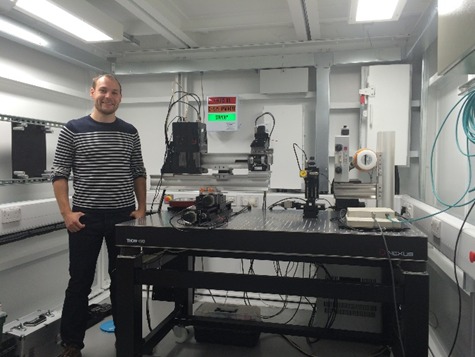
The Colour Imaging Bay is an in-house built system designed as a flexible work bench for spectroscopic X-ray imaging and tomography. In bright field imaging, the energy-sensitive, pixelated detector allows for detection of absorption edges and 3D reconstruction of different atomic elements within a sample. The high-power source (225 kV, 1800 W) allows for X-ray fluorescence (XRF) imaging using a pinhole camera arrangement perpendicular to the beam. X-rays are absorbed and re-emitted from atoms within the sample. These photons have characteristic energies specific to each atomic element. Spatial resolution depends upon the size of the pinhole optic and geometric magnification but is typically in the range of 20 – 200 microns. Scan times can be very long, normally 5 – 10 mins per projection with a full 3D scan taking up to 24 hrs.

The Colour Imaging Bay is under ongoing development and we welcome collaborative proposals for the development.

Key Features
  • In-house system designed as a flexible work bench for spectroscopic X-ray imaging and tomography
  • The spectroscopic capability enables chemical sensitivity combined with imaging in 2D or 3D
  • Reconstructed voxel size 10 µm to 1 mm
  • Linear and rotation motors (various sizes) for flexible experimental set-ups
  • 225 kV, 1800 W X-ray source (Tungsten target)