
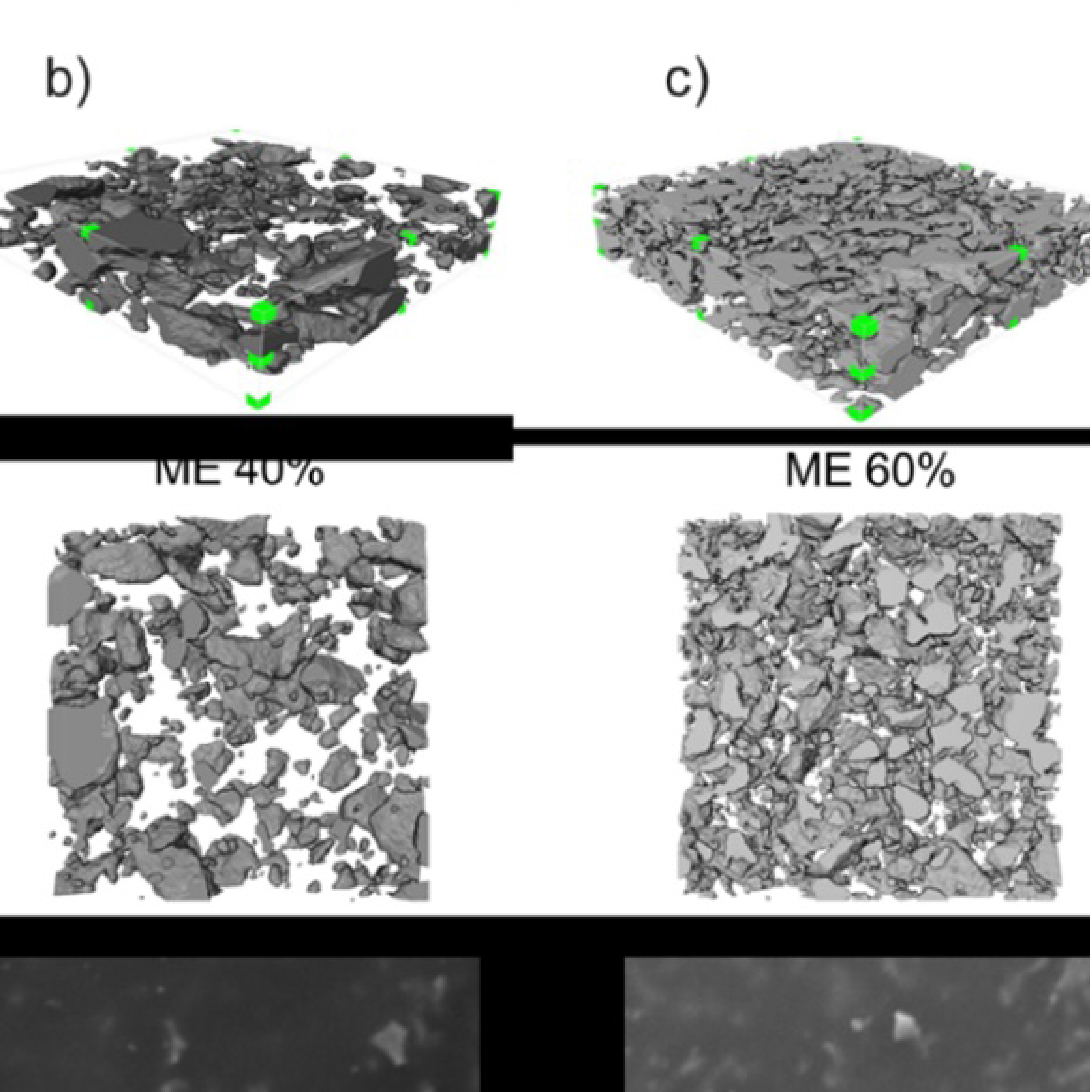
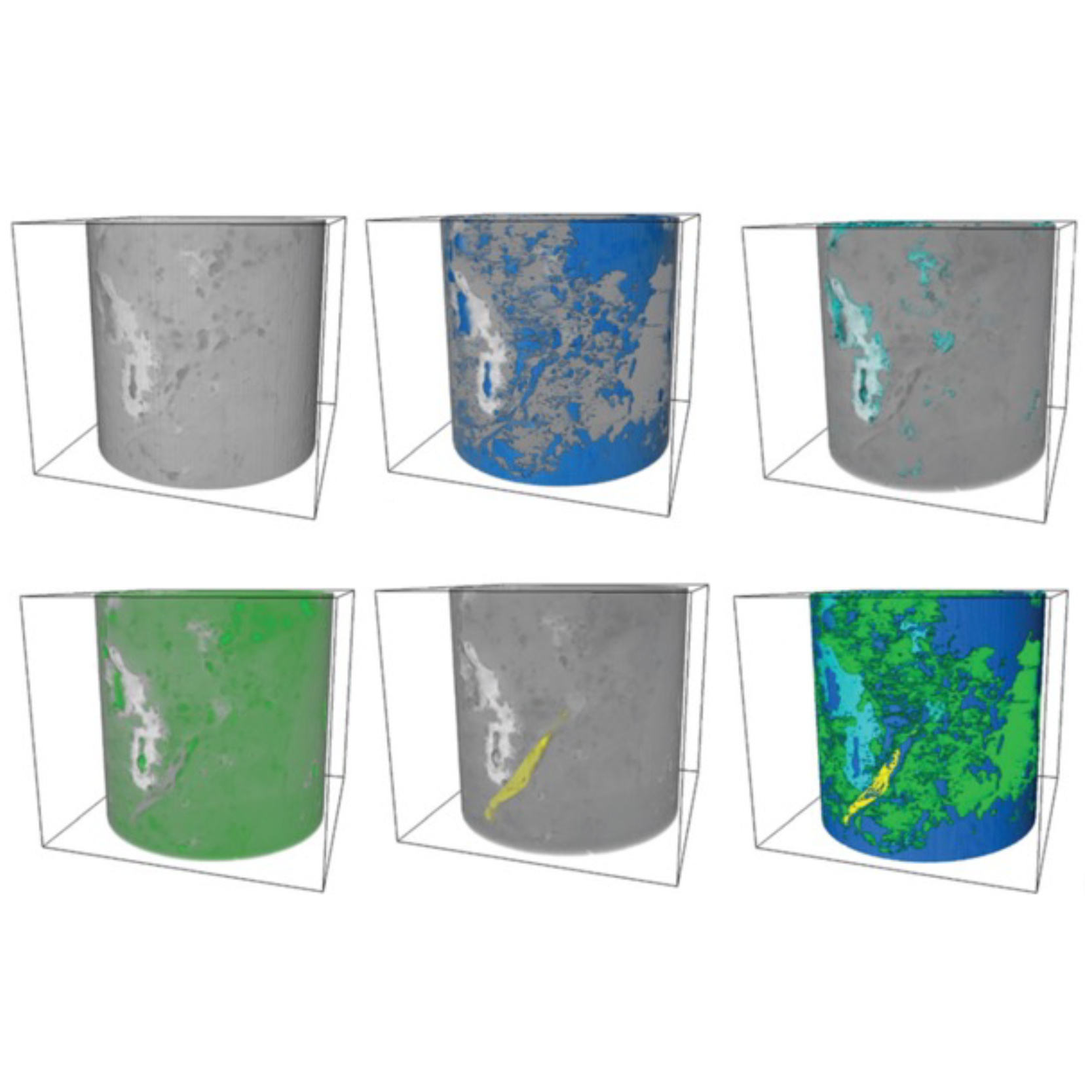
3D Microstructure of Soft Magnetic Elastomer Membrane
Soft magnetic elastomer membranes enable fast magnetic actuation under low fields. In our project, we… Read More
Events & Resources
News, Events and Resources from NXCT Partners
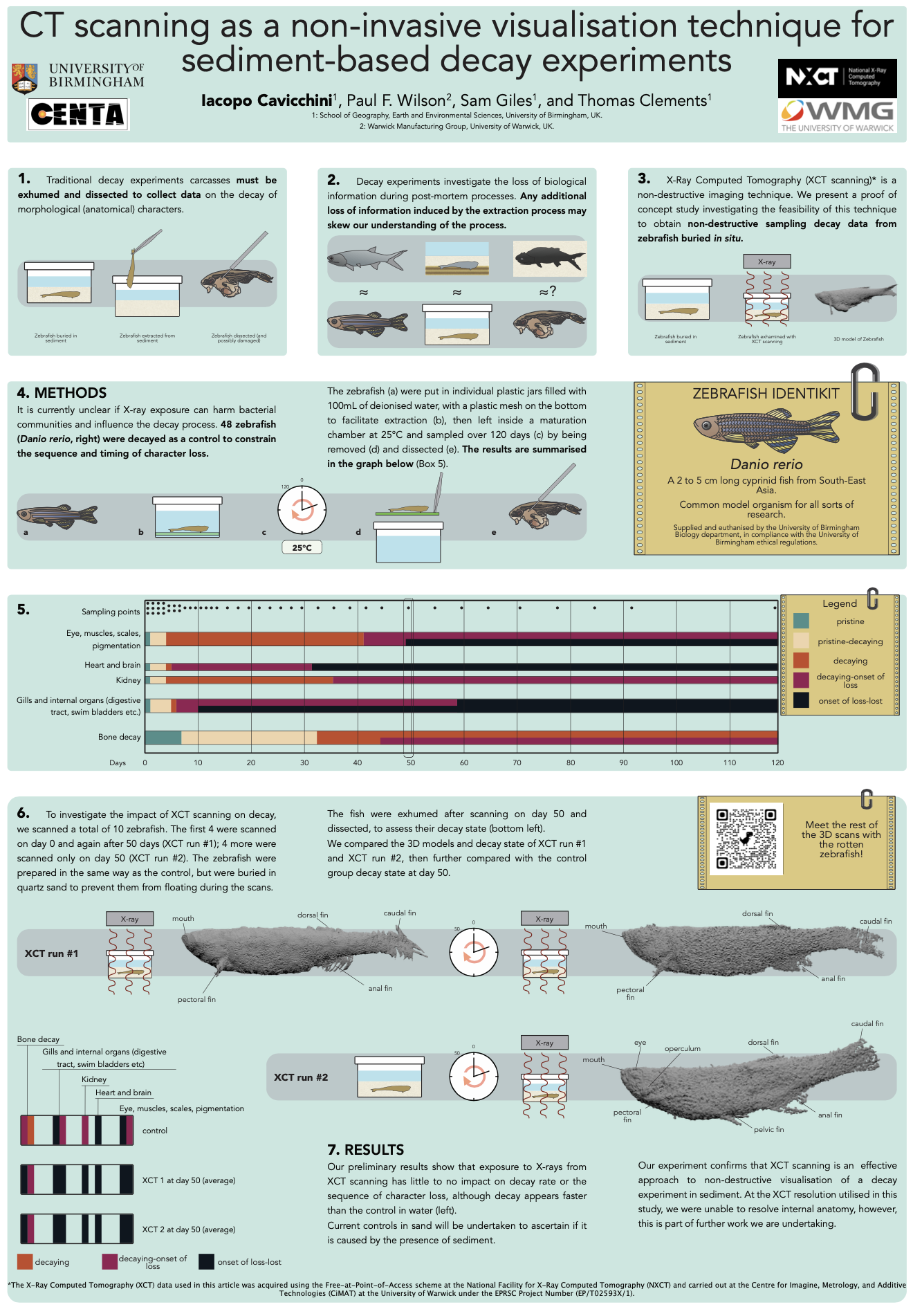
 Understanding the processes of decay is vital to reveal the controls on soft tissue fossilisation. Typically, during decay experiments in sediment, carcasses need to be exhumed during sampling, potentially losing data. We utilised XCT scans to observe a decaying zebrafish entombed within sediment, negating destructive sampling.
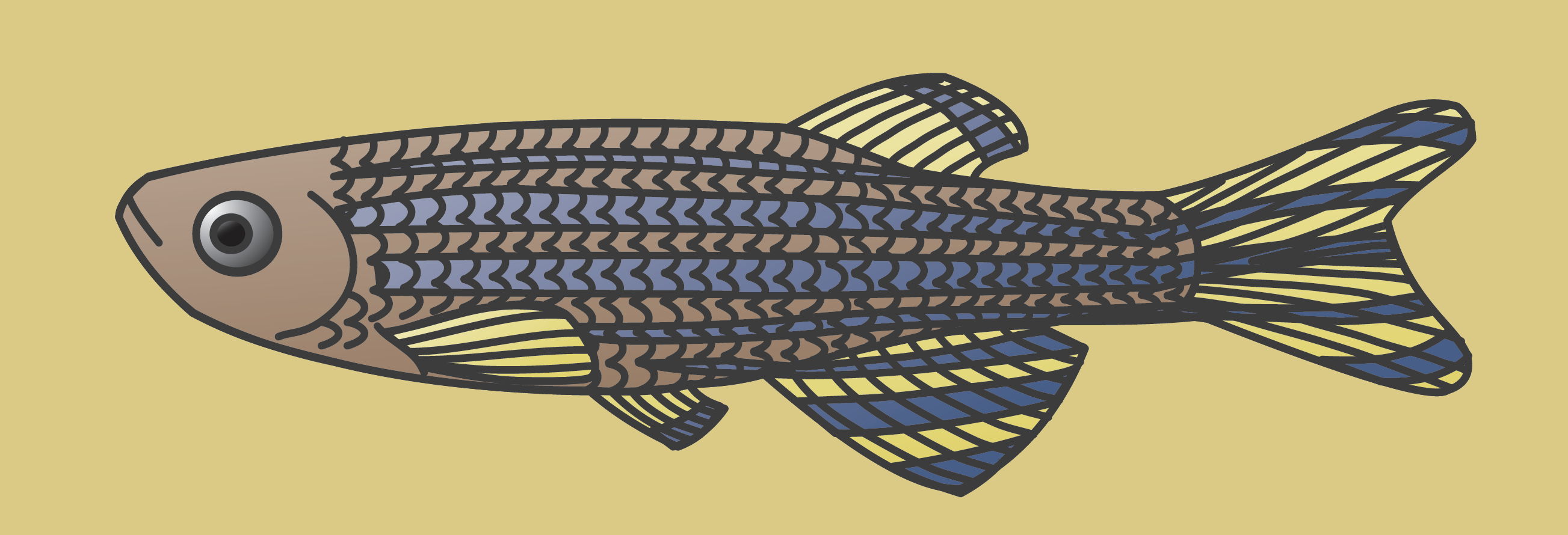
“Tackling taxonomic and taphonomic biases in the Triassic fossil fish record” investigates Early Triassic Fish Fauna species, their fossil record, and fossilisation processes that may bias our understanding of their diversity.
In order to understand the preservation of fish taxa, this project from researchers at the University of Birmingham, utilised decay experiments, investigating the decay of zebrafish in controlled conditions, producing informative data that helps us understand potential preservational biases. However, one issue with decay experiments, especially those in sediment, is that the carcasses need to be exhumed during sampling. Therefore, we proposed to use XCT scans to observe a decaying zebrafish entombed within sediment.
This novel application of XCT scanning is a proof of concept that could alter how taphonomic experiments are undertaken: successful imaging would negate the need to disturb and destructively sample during decay experiments. However, it was possible that CT scanning would destroy or alter the bacterial populations that drive decay, limiting this technique’s usefulness for decay experiments.
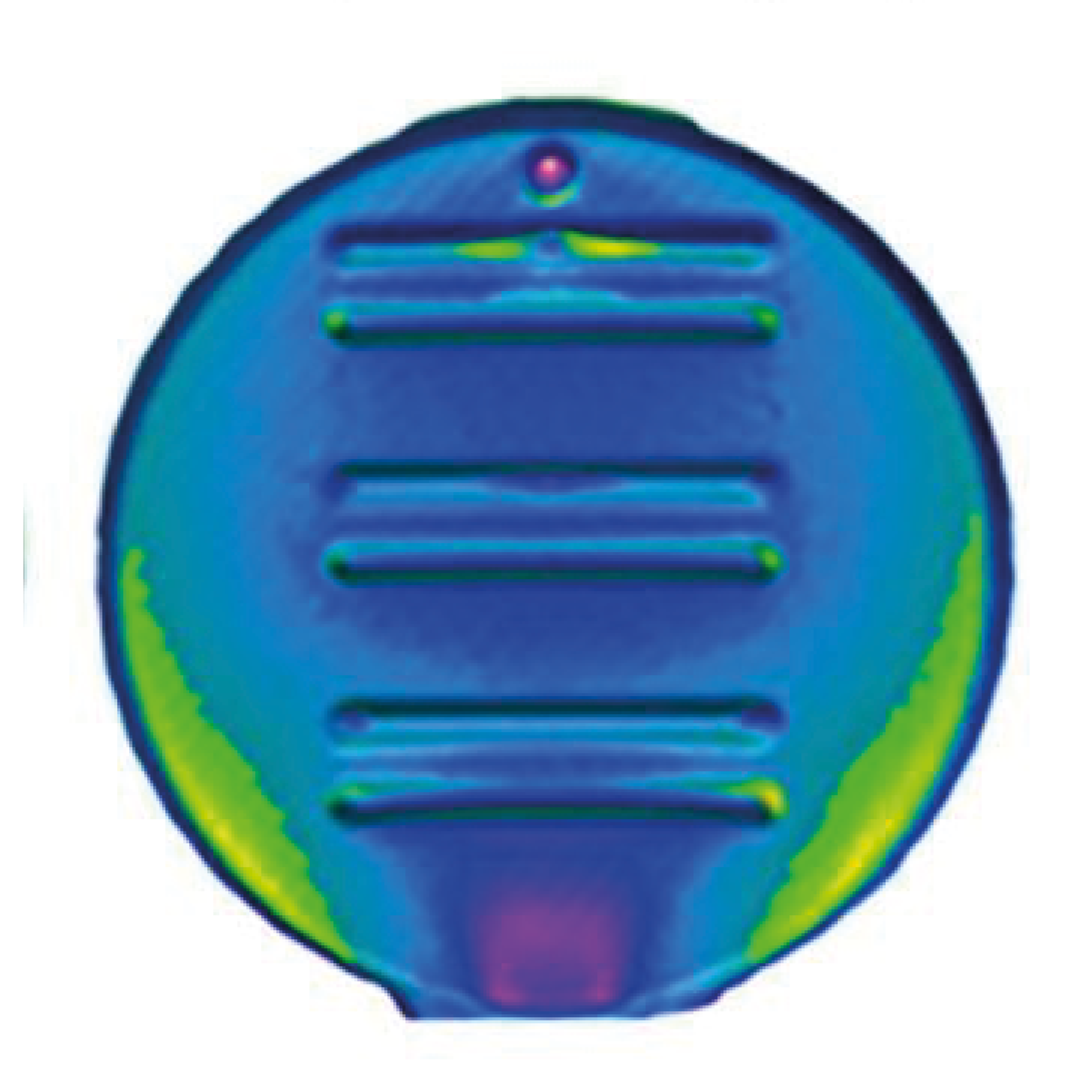
In order to investigate this, the research team buried 8 zebrafish carcasses in sand, and de-ionised water. Four fish were XCT scanned on day one and on day 50 of the experiment. The rest were only scanned day 50. We compared the decay states of the two batches to experiments undertaken in the traditional manner and found no significant difference. The fish carcasses were visible in the XCT images, however, the contrast was not enough to see the internal anatomy, so future work aims at refining the technique.
“This experiment seeks to revolutionise how decay experiments are undertaken, shifting a methodology which stayed unchanged in the last four decades. This work was presented at the annual meeting of the Palaeontological Association in Cork, 2022 (and was awarded the President’s poster prize) and will result in a publication. Furthermore, this work is the basis of continuing experiments investigating using XCT imaging techniques in experimental palaeontology.”
Iacopo Cavicchini, PhD Researcher, University of Birmingham
The XCT scanning was carried out in collaboration with Paul Wilson, University of Warwick, who curated the CT scanning sessions, using the NXCT Free Beamtime scheme at the Centre for Imagine, Metrology, and Additive Technologies (CiMAT) at the University of Warwick under the EPRSC Project Number EP/T02593X/1.
Project Supervisors
Sam Giles, Thomas Clements, Zerina Johanson, Ivan Sansom.
Collaborators
Paul Wilson
Soft magnetic elastomer membranes enable fast magnetic actuation under low fields. In our project, we… Read More
Nowadays, the increasing capability of micro-manufacturing processes enables the manufacture of miniature products with extremely… Read More
Injection of CO2 into shale reservoirs to enhance gas recovery and simultaneously sequester greenhouse… Read More
