
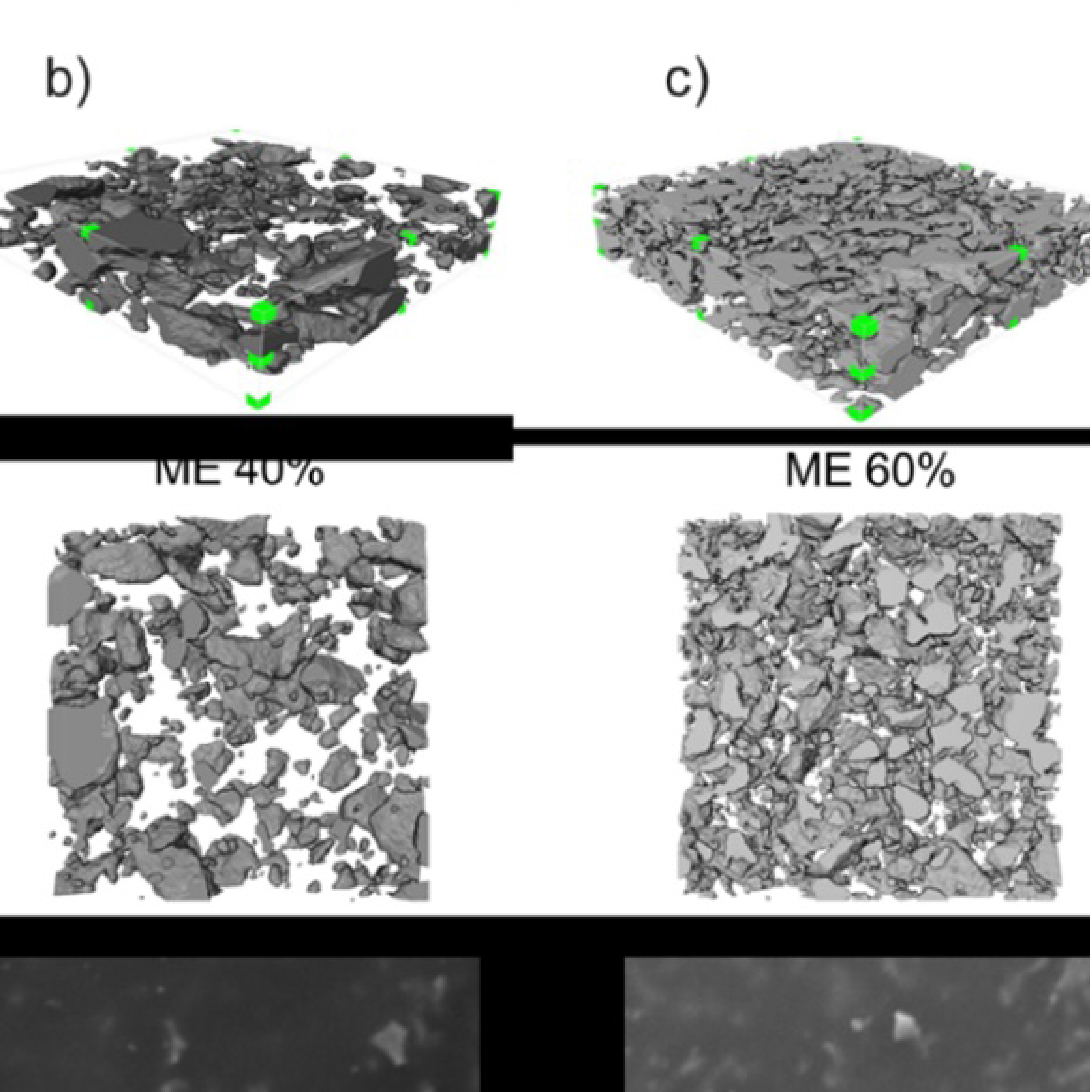
3D Microstructure of Soft Magnetic Elastomer Membrane
Soft magnetic elastomer membranes enable fast magnetic actuation under low fields. In our project, we… Read More
Events & Resources
News, Events and Resources from NXCT Partners
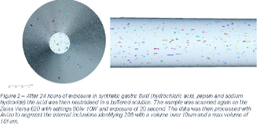
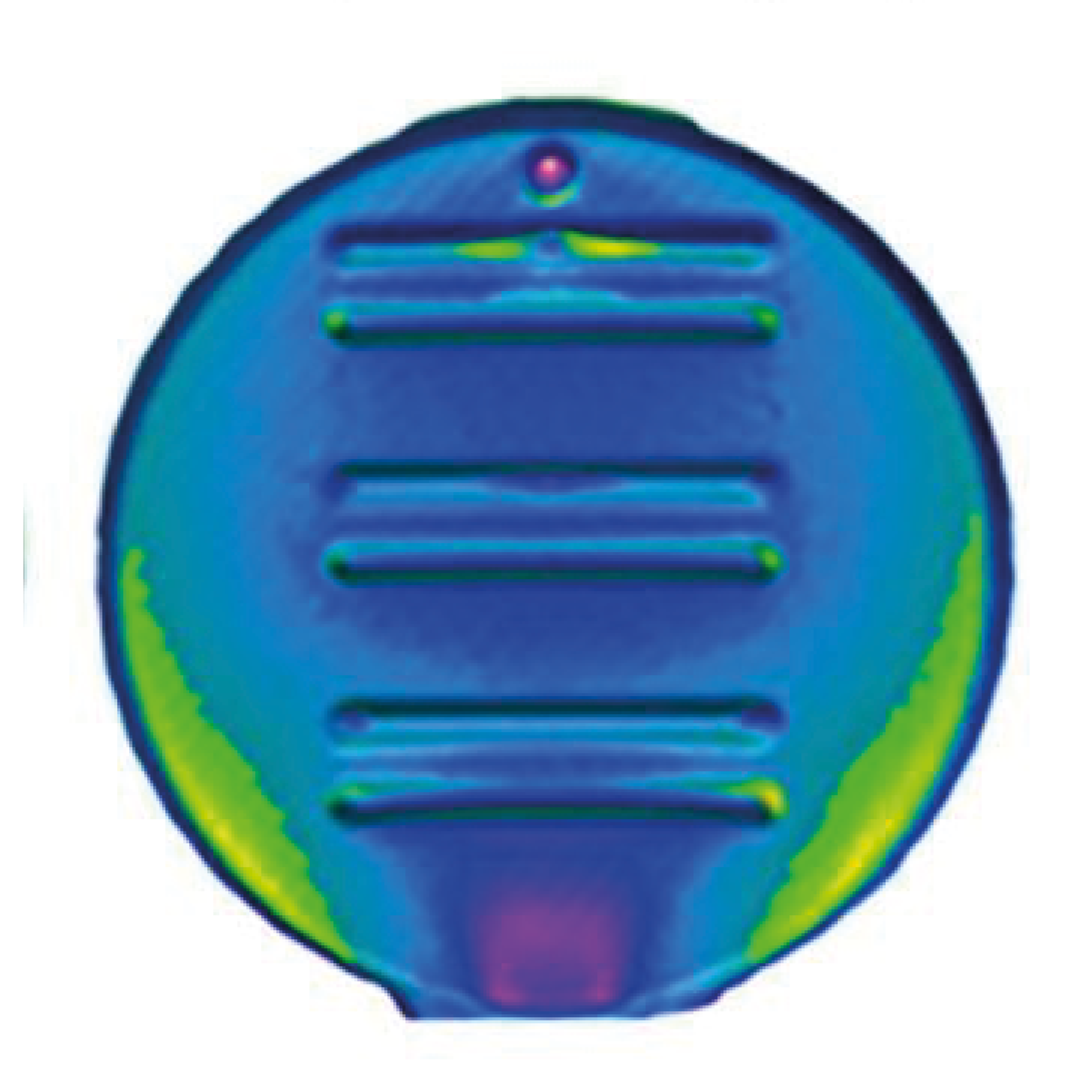
Nitinol is a common metal which is used in several medical devices, in particular this material can be found in oesophageal stents as a palliative care device allowing for a closed off oesophagus to be opened and allow for food and liquid to pass. At present, these stents are only in service for 4 to 6 months in service before requiring intervention due to device failure or migration. Through the use of CT it has been possible to show the effects that the gastric fluid had on the internal structure of the metal.
A common form of cancer that accounts for 5% of all cancer diagnoses worldwide is oesophageal cancer which commonly causes dysphagia, a blockage which stops food and drinks from freely passing. The traditional treatment for this symptom is the use of a stent which pushes the intruding tumour aside allowing food and drink to freely pass. These stents can come in a variety of materials but are often made of nitinol. In recent years a trend has been noticed around the failure of these stents while in service requiring removal and replacement. Understanding the failure of these devices is needed to increase the longevity of these devices improving quality of life for the patients.
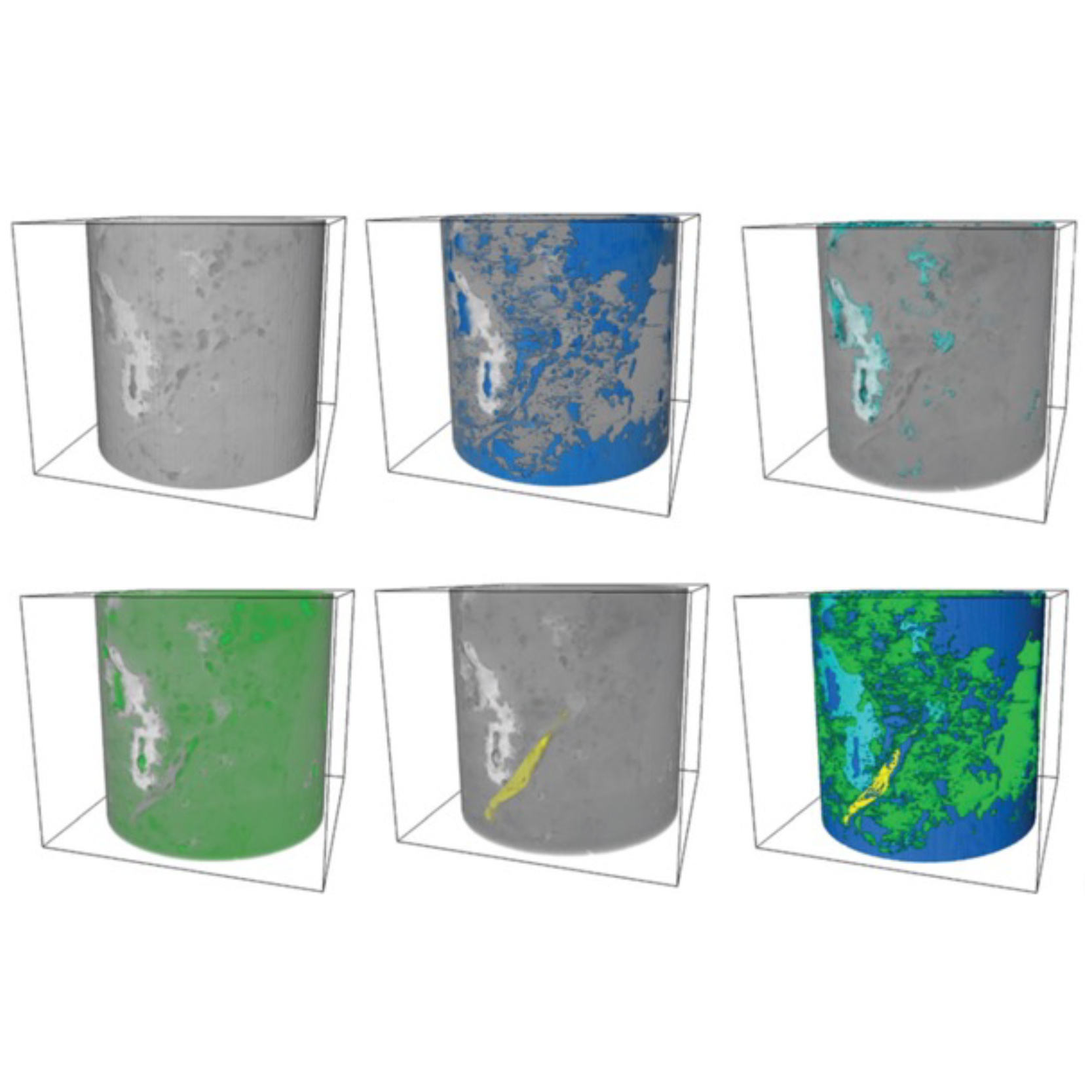
The project completed with the aid of the NXCT was to look at the embrittlement process while immersed in synthetic gastric fluid over 24, 8, 4, 2, 1 and 0.5 hours. 5mm of 0.25mm diameter nitinol wire was mounted using epoxy on a aluminium rod. A first scan was completed to take the start state of the wire then further scans were completed after each period of immersion. After immersion the wire was placed in the Zeiss Versa 620 with 20x optical magnification. The output of this project was the ability to identify the inclusions within the material and the development of further inclusions dur to embrittlement which can be seen increasing in size and number after each immersion step.
The knowledge provided and access to the equipment by the NXCT has helped get the best quality images and ensure that the data gathered was as similar as possible during each scan. In addition, the NXCT helped process the data and allowed me to get the most from it.
This work has the potential to show that even over a short period of time that the embrittlement while in the gastric fluid can take effect. Further work is planned to determine the effects that this embrittlement has on the failure of the material with the potential to use CT to try and identify the initiation point.
“This work is significant as it has allowed us to create a time lapse of the embrittlement process and see what impact it is having on the internal structure the metal. This is the first time that we can see the effect that embrittlement has on the internals structure over time.”
Ryan Weller, Postgraduate researcher

Soft magnetic elastomer membranes enable fast magnetic actuation under low fields. In our project, we… Read More
Nowadays, the increasing capability of micro-manufacturing processes enables the manufacture of miniature products with extremely… Read More
Injection of CO2 into shale reservoirs to enhance gas recovery and simultaneously sequester greenhouse… Read More
