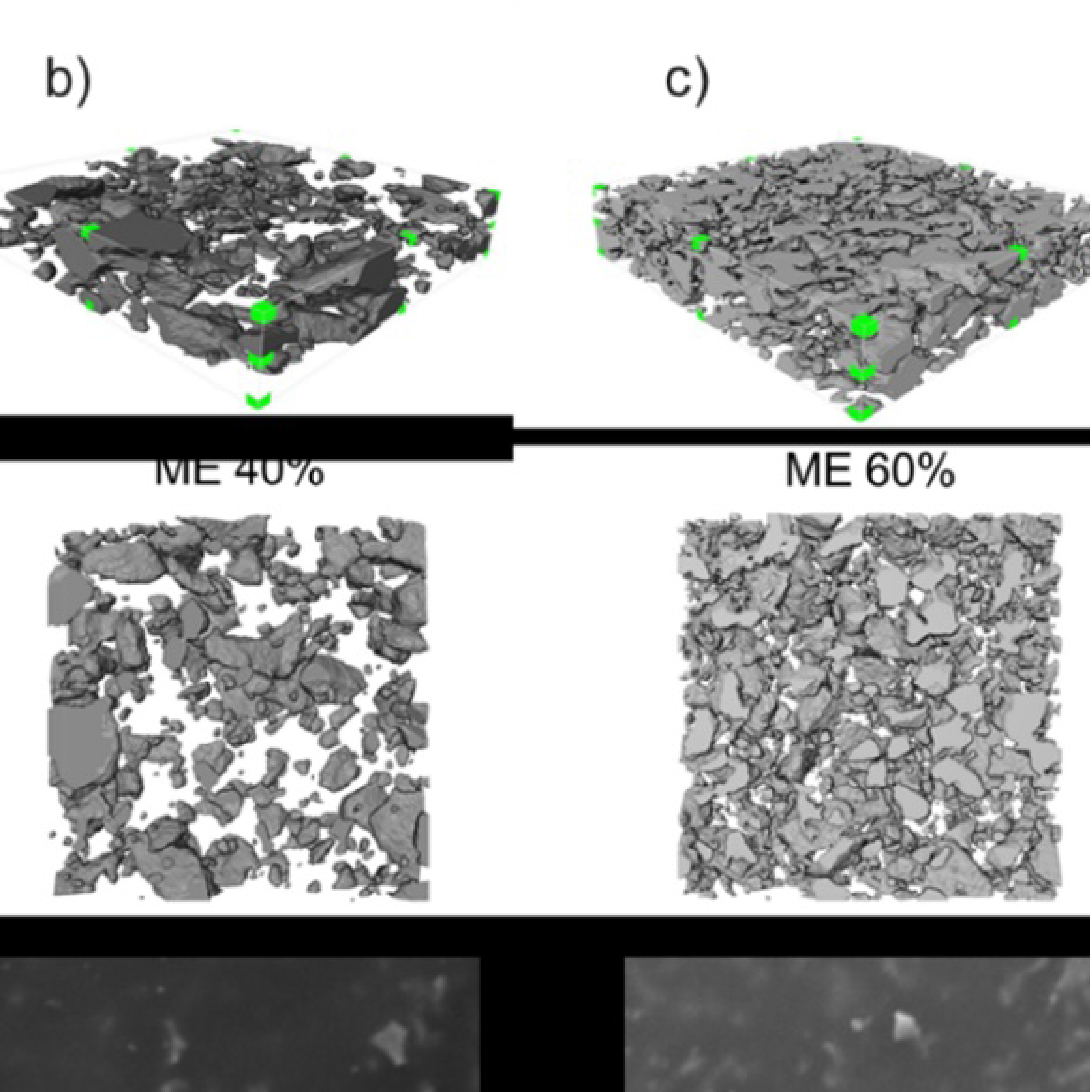
3D Microstructure of Soft Magnetic Elastomer Membrane
Soft magnetic elastomer membranes enable fast magnetic actuation under low fields. In our project, we… Read More
Events & Resources
News, Events and Resources from NXCT Partners
A phage cocktail targeting E.coli biofilms in catheter-associated infections achieved over 97% biofilm reduction, as shown by detailed CT scans using the Tescan Unitom XL. These findings highlight phages’ potential as a promising alternative to antibiotics for treating resistant bacterial infections and improving patient outcomes.
Escherichia coliis responsible for the majority of cases of both urinary tract infections (UTIs) and bloodstream infections (BSIs). UTIs which spread into the bloodstream causing BSIs–cases known asurosepsis–account for approximately 25% of all BSIs in adults and 5% of severe BSIs and septic shock. In the UK, the mortality rate for BSIs with treatment is approximately 25%-#1 cause of in-hospital death. The emergence of antibiotic-resistant strains of this species has become a growing concern in healthcare, prompting the search for alternative treatments; bacteriophages, viruses which infect and kill bacteria, are one such alternative.In order to test the effect of phages on robust, patient-derived infections, a three-phage cocktail was created, based on in vitro assays. Catheters were sourced from long-term catheterised patients (>8 weeks) with symptoms of UTIs, and cultured to identify those containingE. coli within the polymicrobial infections (n = 4).Each catheter was cut into two, with one segment placed in the fridge to act as an untreated baseline, and the other segment treated in an Artificial Urinary Tract model developed to simulate a 5-day intravenous administration of phages, where the catheters simulate urinary tracts with the presence of infection. After the phage administration, the baseline and treated segments were scanned on a Tescan Unitom XL atCiMAT, University of Warwick. Residual biofilms were calibrated using a density phantom and so had theirdensity, volume, and thickness quantified; the ability to provide quantitative as well as qualitative information is an improvement on the microbiological gold standard method of electron microscopy. The CT scans show that the phage cocktail had a profound effect on the catheter biofilms: all catheters displayed a considerable decrease in biofilm volume, with a >97% reduction in three of the four; all four also had a considerable decrease in biofilm thickness, with two displaying a >92% reduction in average thickness and>93% reduction in maximum thickness. With further work, these findings could contribute to the development of more effective treatments for antibiotic-resistant bacterial infections and the understanding of the effect of phages on biofilms, and ultimately form the basis for creating phage cocktails for clinical use.
This is the first study of its kind in demonstrating the treatment with bacteriophages of ex vivo patient-derived biofilms,andis also the first study involvingimaging and quantification ofmedicalbiofilms usingmicro-CT. The outstanding results of the phage experiments and the detailed information from the CT scanspaint a clear picture that paves the wayto further understanding of biofilms, catheter-associated urinarytract infections, and phage treatment of polymicrobial infections.
Karen AdlerPhD StudentLeicester Centre for Phage ResearchDepartment of Genetics, Genomics and Cancer Sciences University of Leicester
Collaborator: Paul Wilson, University of Warwick (CiMAT)


Soft magnetic elastomer membranes enable fast magnetic actuation under low fields. In our project, we… Read More

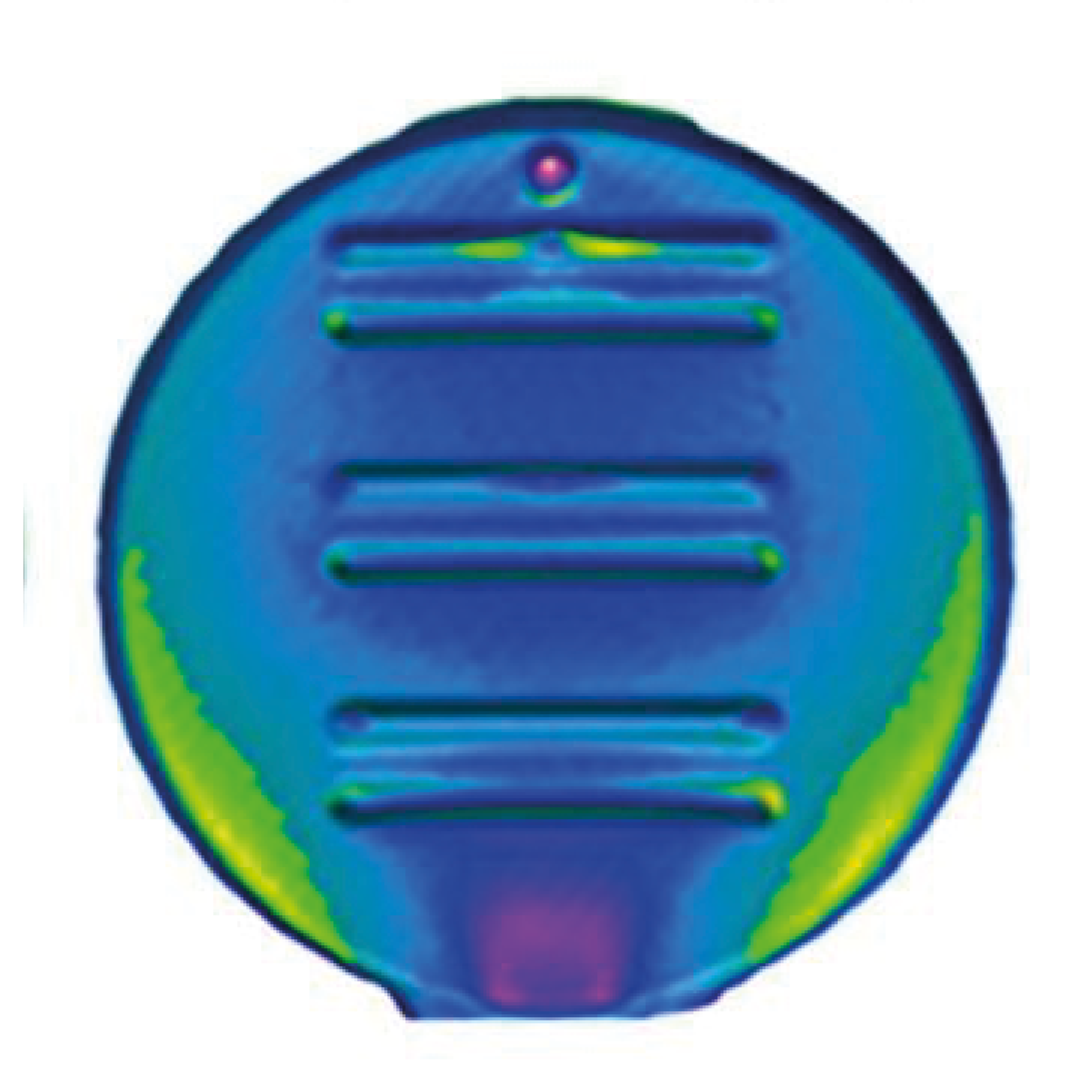
Nowadays, the increasing capability of micro-manufacturing processes enables the manufacture of miniature products with extremely… Read More

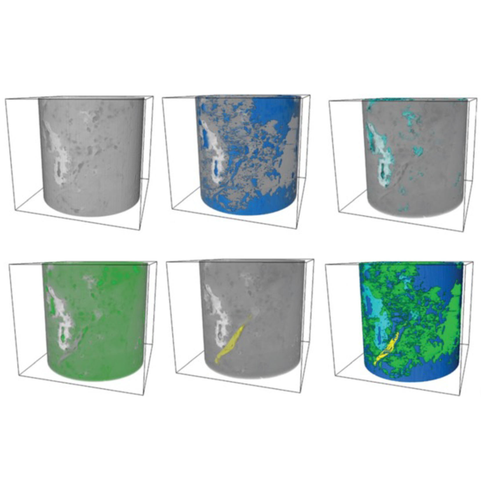
Injection of CO2 into shale reservoirs to enhance gas recovery and simultaneously sequester greenhouse… Read More