
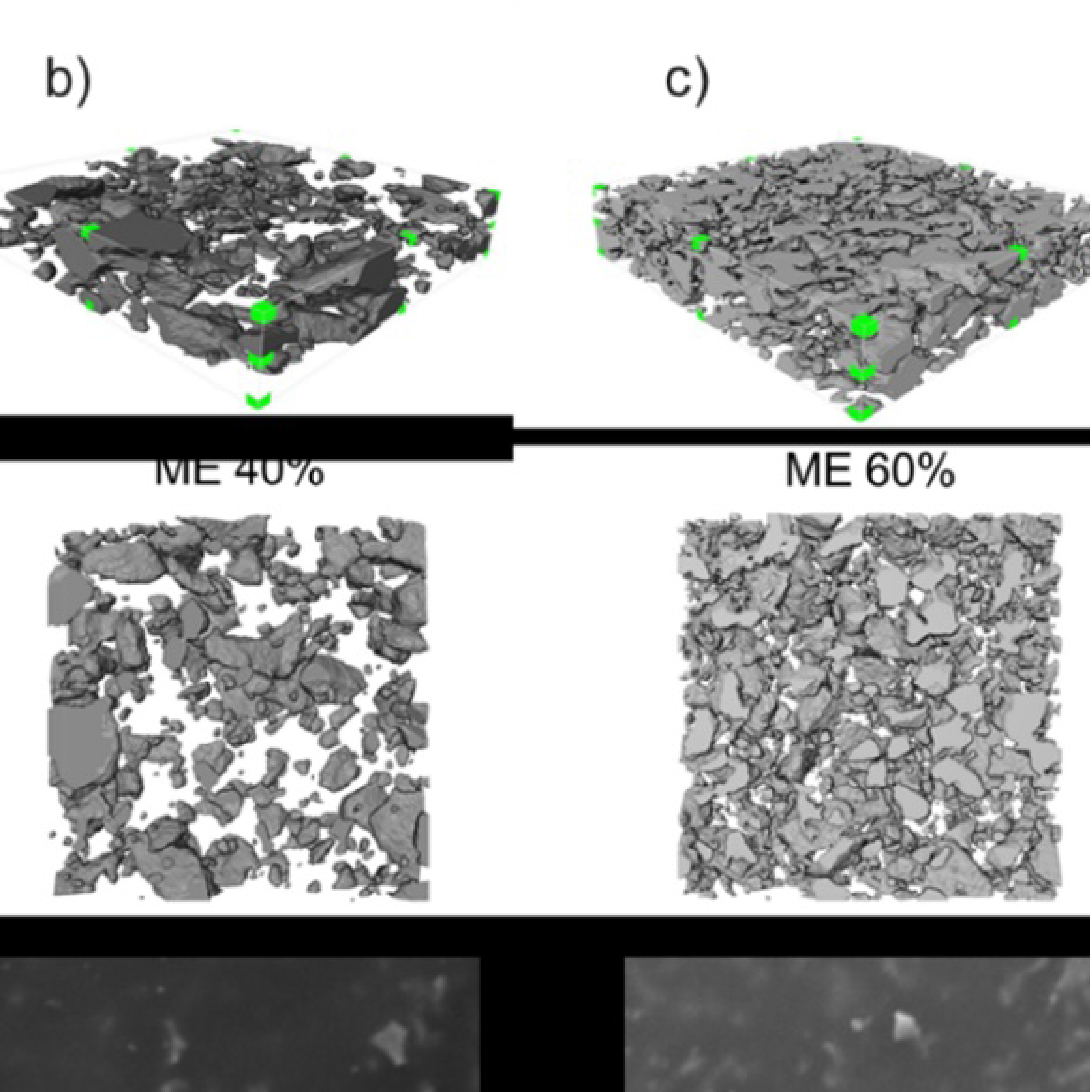
3D Microstructure of Soft Magnetic Elastomer Membrane
Soft magnetic elastomer membranes enable fast magnetic actuation under low fields. In our project, we… Read More
Events & Resources
News, Events and Resources from NXCT Partners
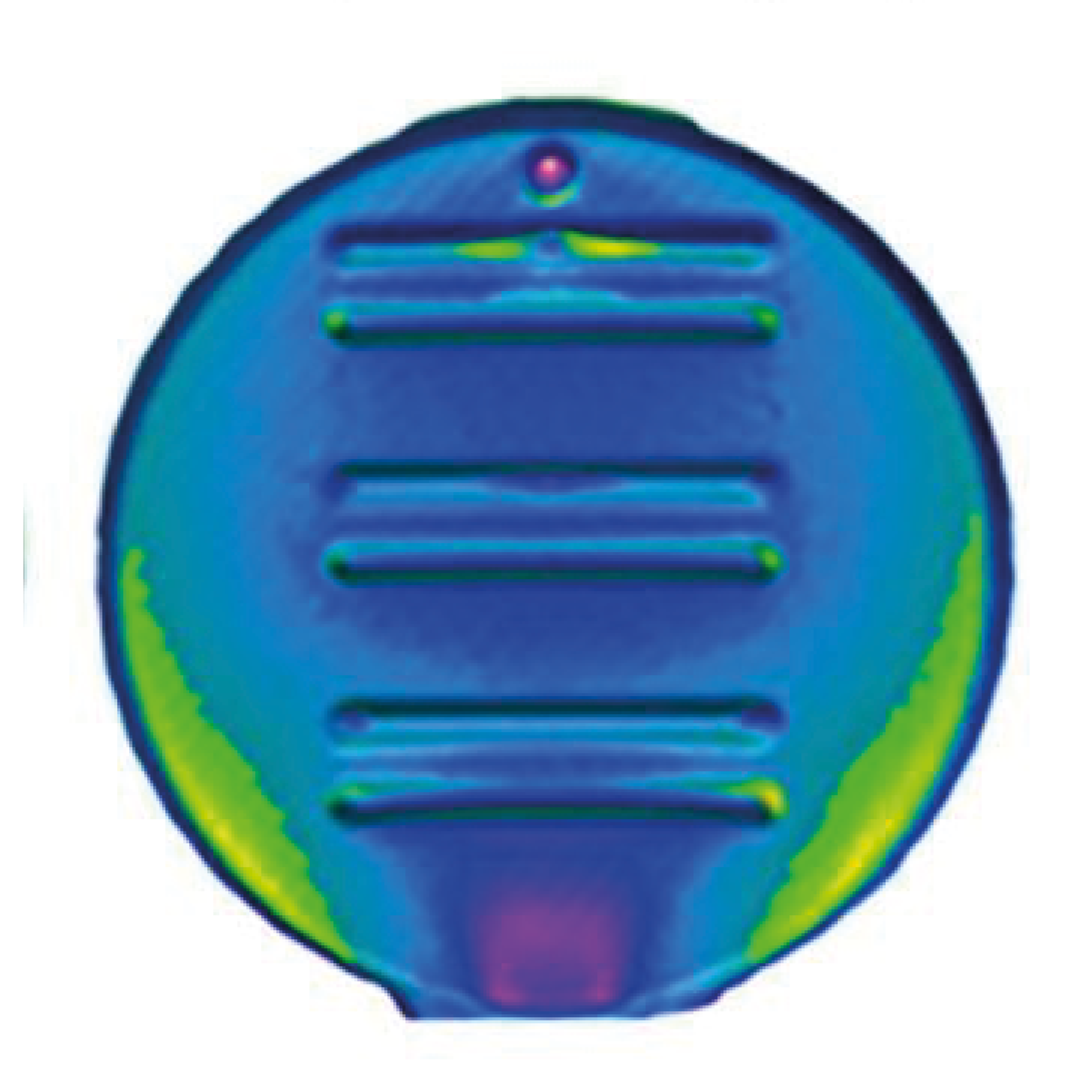
Conventional x-ray imaging is useful for separating materials according to their density or composition, based on differing levels of attenuation. However, this is averaged within a voxel, and information on the nature of the microstructure is lost. This is where dark-field imaging is powerful, as contrast is generated from sub-voxel features such as pores, fibres, or cracks. Dark-field tomography can thus build three-dimensional volumes that show the microstructure of the material.
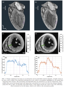
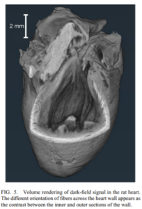
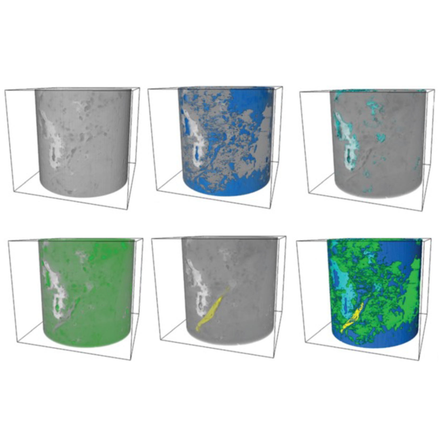
One of the machines available through the NXCT is the edge-illumination x-ray imaging system. This system has sensitivity to the dark-field signal and can measure the dark-field and attenuation signals simultaneously for a complimentary set of images. After developing the theory behind how dark-field volumes can be reconstructed from a series of projections, we took a number of scans of different samples using the edge-illumination system through the NXCT. These included a short-fibre reinforced composite, pouch cell battery, tissue-engineered oesophagus, and a rat model heart (pictured).
One particularly interesting result came from the rat heart sample. After reconstructing the attenuation and dark-field volumes, we saw significant contrast within the heart wall in the dark-field slices, something not seen in the corresponding attenuation slices. After further investigation we concluded that this contrast originated from the fibres within the heart wall structure at differing orientations. The fibres within the middle of the heart wall were orientated circumferentially around the ventricle, but those fibres towards the inner and outer surfaces are aligned vertically, from the apex to the base.This sensitivity to fibre orientation is only possible due to the microstructural sensitivity available with dark-field contrast.A second result was that the dark-field reconstruction was sharper by roughly 50% when compared to the attenuation reconstruction. This is due to boundaries showing a high dark-field signal, and thus the edges of the sample were strongly defined.
Going forward, we believe that dark-field tomography will be extremely useful for investigating the microstructure of samples, giving complimentary information to the conventional attenuation volumes. This is particularly useful for porous samples such as lungs or bone and could improve the capability to detect cracks or defects in composite materials. The availability to do so with a laboratory-based system, one that isalso offered through the NXCT, means that this can be done without the need of travelling to highly brilliant synchrotron sources.
“Edge-illumination x-ray dark-field tomography allows us to image sub-voxel pores and fibres within soft tissues and materials, with complimentary contrast to conventional attenuation imaging, which has great potential for improving diagnostic accuracy and aiding in materials science research. I believe x-ray dark-field tomography will enable us to explore the microstructure of new materials and I am looking forward to seeing the ways in which this technology will be utilized in the future.”
Adam Doherty
Research Assistant
Advanced X-ray Imaging Group (AXIm)
Department of Medical Physics and Biomedical Engineering, University College London
Malet Place Engineering Building Room 2.20

Soft magnetic elastomer membranes enable fast magnetic actuation under low fields. In our project, we… Read More
Nowadays, the increasing capability of micro-manufacturing processes enables the manufacture of miniature products with extremely… Read More
Injection of CO2 into shale reservoirs to enhance gas recovery and simultaneously sequester greenhouse… Read More
