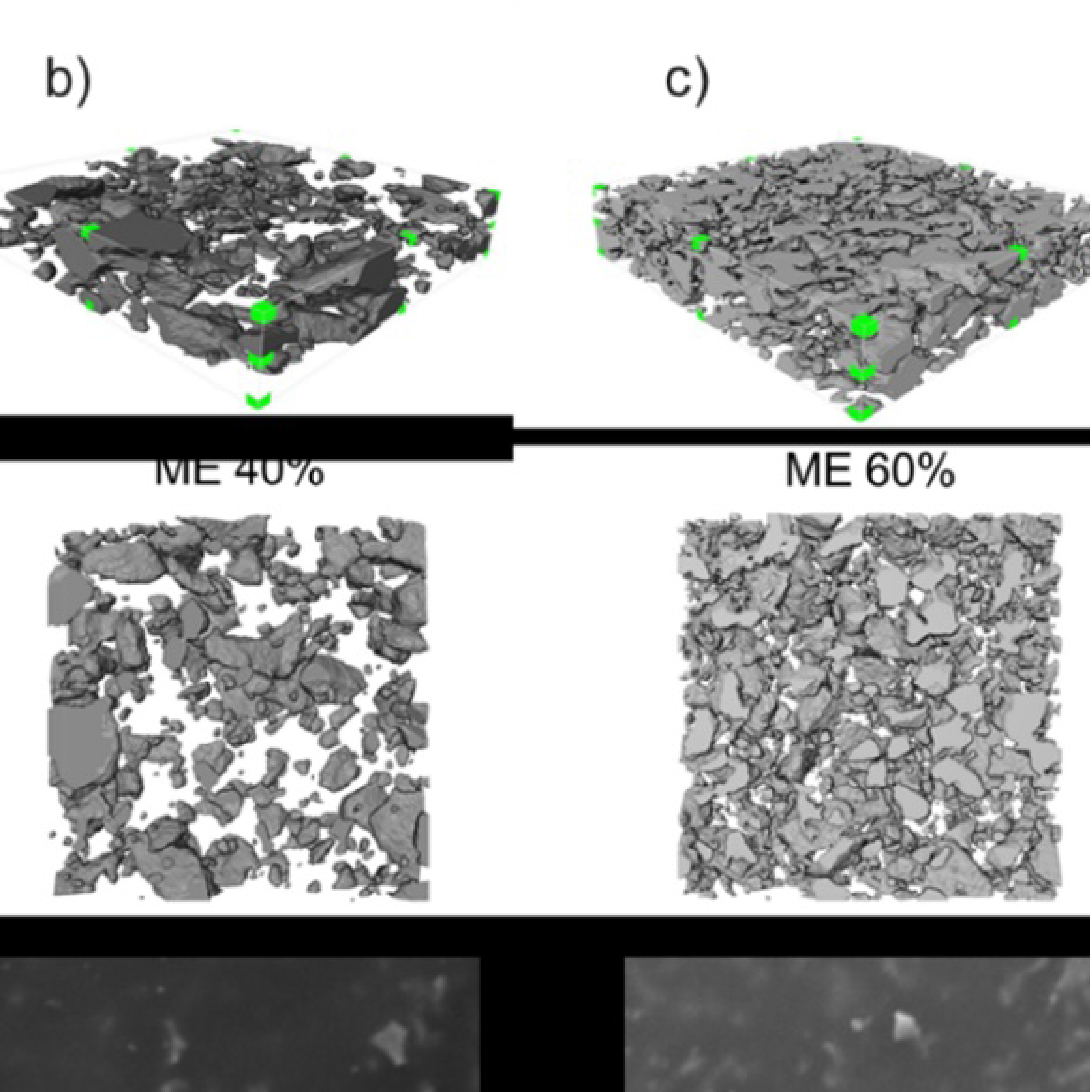
3D Microstructure of Soft Magnetic Elastomer Membrane
Soft magnetic elastomer membranes enable fast magnetic actuation under low fields. In our project, we… Read More
Events & Resources
News, Events and Resources from NXCT Partners
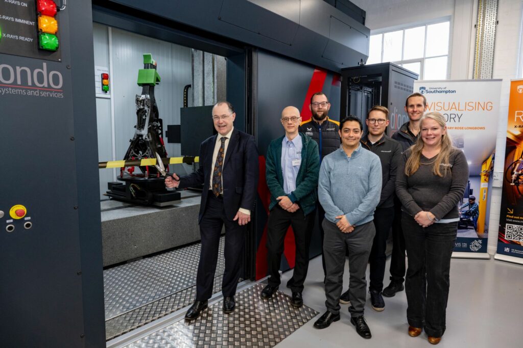
This week the University of Southampton opened a new x-ray scanner, the largest of its type within a UK University! The diondo d5 takes samples up to 200kg in weight and 2.2m in height, and increases our ability to examine even large items.
The Diondo d5 system is a walk-in scanner funded by the EPSRC as part of the The National X‑ray Computed Tomography facility (NXCT; nxct.ac.uk) infrastructure development at the University of Southampson.
The d5 is equipped with two X-ray sources: one 300 kVp micro-focus transmission tube enabling higher spatial resolution (<5 µm) scanning of larger/denser objects and one high-energy 450 kVp mini-focus reflection tube offering higher X-ray flux for faster, lower noise scanning. It has a customisable 3000 x 3000 pixels Flat Panel (FP) detector (active area 417 x 417 mm) allowing the operator to optimise the system’s performance to best suit the sample and imaging requirements. The system it is also equipped with a range of advanced acquisition modes which allow horizontal and vertical field of view expansion, including helical acquisition, panel offsetting, and vertically overlapping scans. It also features scatter compensation techniques for denser objects and linear translation laminography for high aspect ratio samples.
The d5 can be used to image specimens with dimensions from a few millimetres in cross-section up to objects with ~ 2 –3 m linear dimensions with a scannable field-of-view in excess of 1 m (Ø) x 1.5 m (H). The heavy-duty rotation stage can support specimens up to 200 kg. A labyrinth at the rear of the system allows routing of cables and pipes that can control and feed in situ rigs for time-resolved and/or environment controlled µ-CT experiments.
‣ 300 kVp & 450 kVp X-Ray sources
‣ resolutions:
‣ ‣ 300 kVp source: ~ 3 µm up to 10 W / 20 µm up to 80W
‣ ‣ 450 kVp source: ~ 0.4 mm up to 700 W / 1 mm up to 1500 W
‣ 3000 x 3000 FP detector (active area 417 x 417mm)
‣ scanning modes:
‣ ‣ horizontal & vertical field-of-view expansion and cropping
‣ ‣ helical scanning
‣ ‣ linear translation laminograpgy
‣ scattering compensation
‣ imaging volume in excess of 1x1x1.5 m
‣ 200 kg rating sample stage
‣ temperature controlled
‣ user labyrinth and large 5 x 3.5 x 3.5 m enclosure for large specimens and/or complex in situ testing


Soft magnetic elastomer membranes enable fast magnetic actuation under low fields. In our project, we… Read More

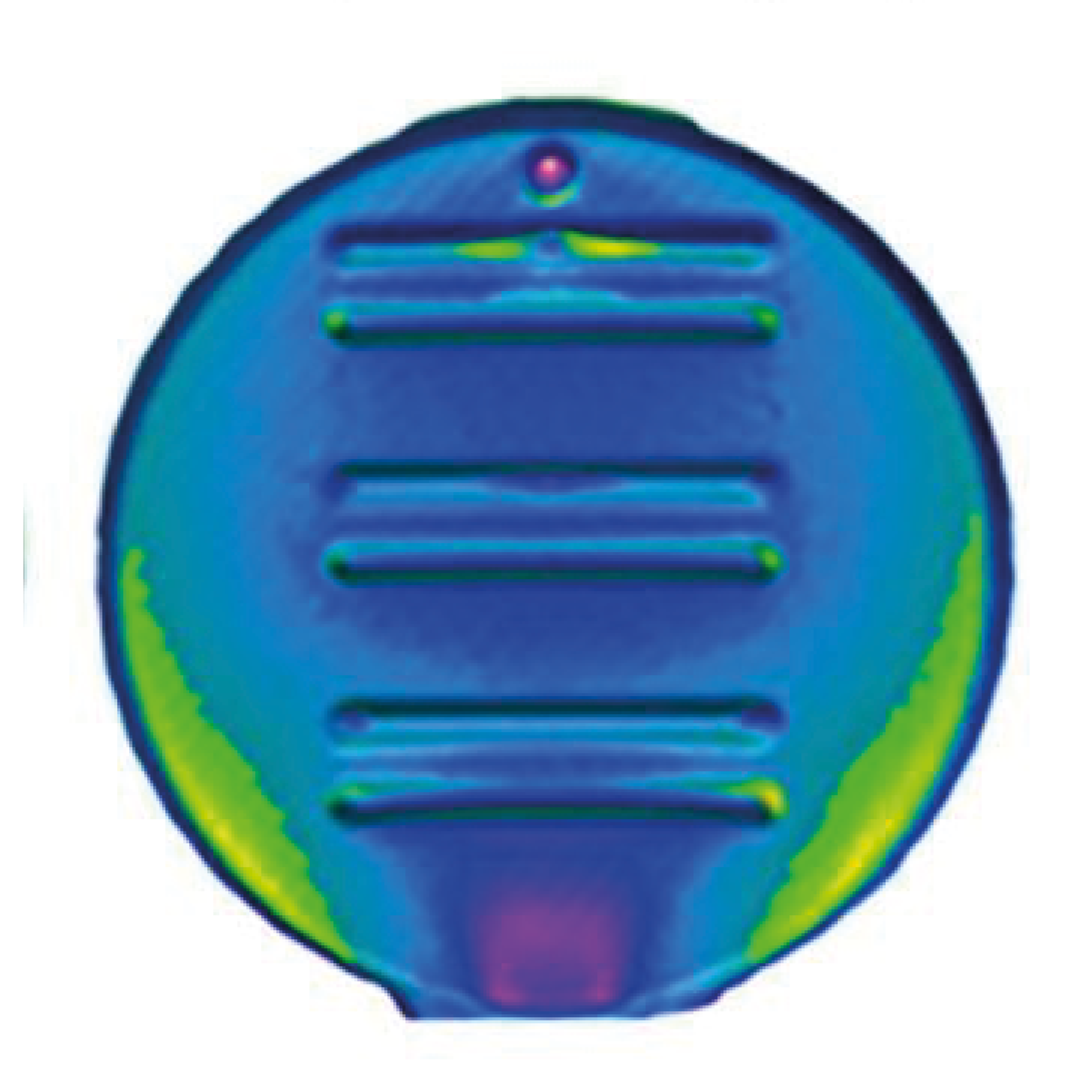
Nowadays, the increasing capability of micro-manufacturing processes enables the manufacture of miniature products with extremely… Read More

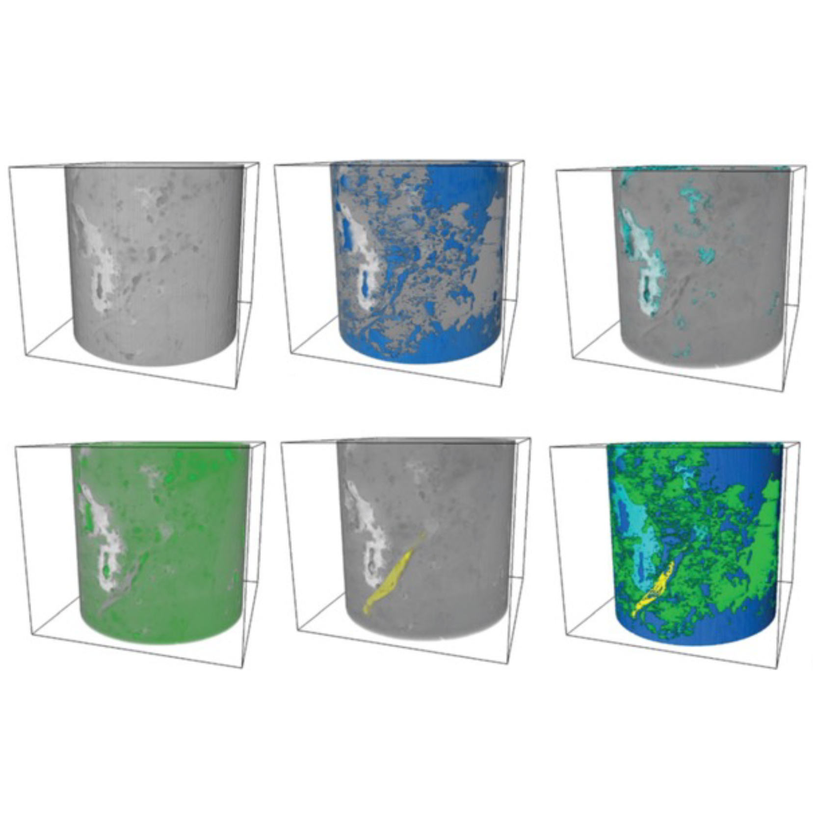
Injection of CO2 into shale reservoirs to enhance gas recovery and simultaneously sequester greenhouse… Read More