
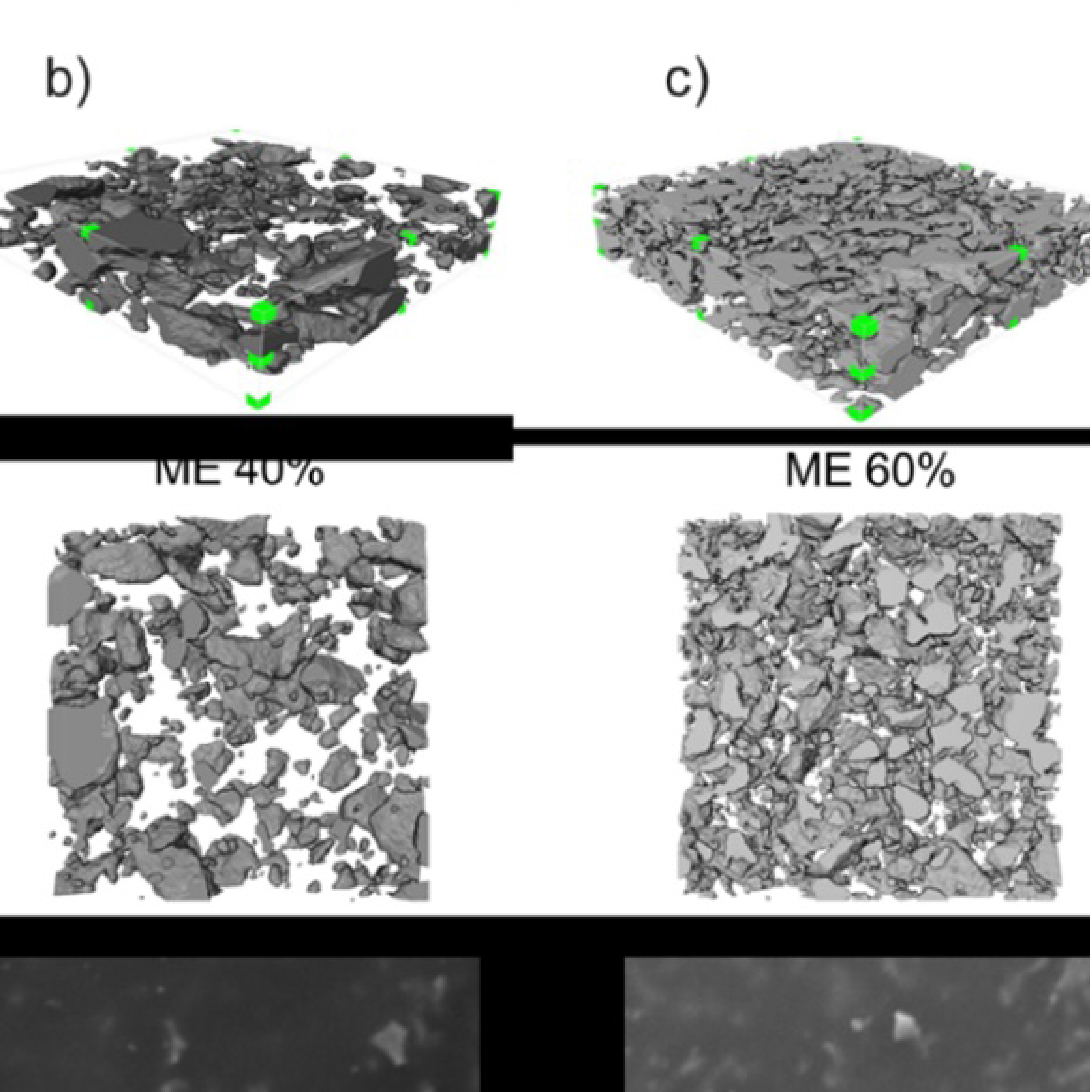
3D Microstructure of Soft Magnetic Elastomer Membrane
Soft magnetic elastomer membranes enable fast magnetic actuation under low fields. In our project, we… Read More
Events & Resources
News, Events and Resources from NXCT Partners
The aim of this project is to exploit these new experimental capabilities to study novel polymeric and hybrid-organic materials that have promising applications as drug carrier and tissue engineering.
Examples such as polyimide (PI) and polyhydroxyalkanoates (PHAs)show adapted capability as monomers of molecular chains can easily be modified and compared with conventional liposome, they demonstrate outstanding biocompatibility, nontoxicity, self-assembly, more drug loading and targeting property after modification.
For instance, modifying with hydrophilic groups such as PEG, materials trend to be self-assemble and form vesicle structures. Loads of assays have been carried on analyzing the effect of vesicles for drug delivery and progresses have been made towards their biomedical applications. But technically speaking, like the lipid bilayer structure, the bilayer structure of vesicle is also ideal approximate assumption. In fact, the asymmetric bilayer structure is inserted with asymmetric protein as receptors. However, little is known about the structures of vesicle membranes and little researches were focused on the physical interactions between these nanomaterials and biological systems. With the restriction of relatively low spatial resolution structure mapping and the complexity of specimen making for CT in the past, researchers usually can obtain only outlines of vesicles with relatively little information, particularly regarding the interactions between materials. But with the help of nano-CT with higher spatial resolution nanoscale structural and composition information could be extracted, and with the help of other electron microscopy elemental mapping, chemical compositional information could be studied to have a deeper understanding of biomaterials.
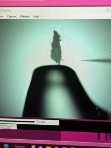
“Micro- to Nano-scale CT would fill out the resolution gap between the general optical microscopes and electron microscopes with its ability to have the 3D reconstruction of my silk fibroin sample. And besides, due to the non-destructed nature of X-ray scan, the same piece of silk fibroin sample can be used to do a series of top-down imaging from optical microscope to electron microscope to extract micro- to nano-scale information.”
Bing Han PhD at the University of Manchester
Soft magnetic elastomer membranes enable fast magnetic actuation under low fields. In our project, we… Read More
Nowadays, the increasing capability of micro-manufacturing processes enables the manufacture of miniature products with extremely… Read More
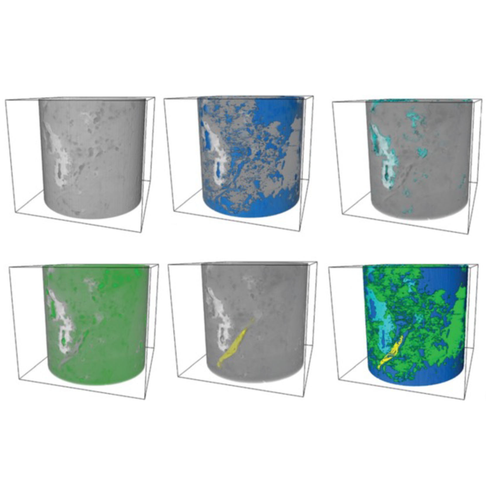
Injection of CO2 into shale reservoirs to enhance gas recovery and simultaneously sequester greenhouse… Read More
