
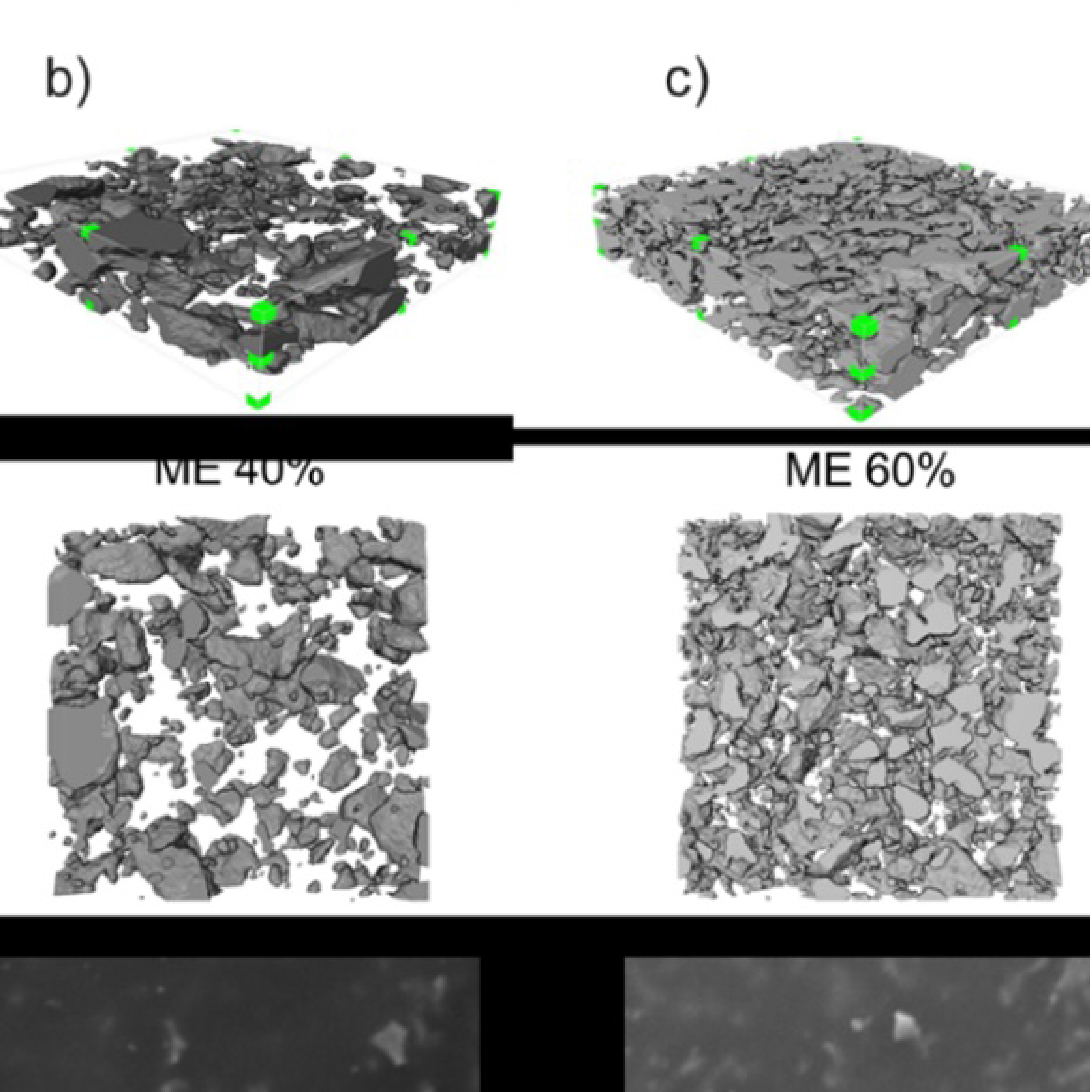
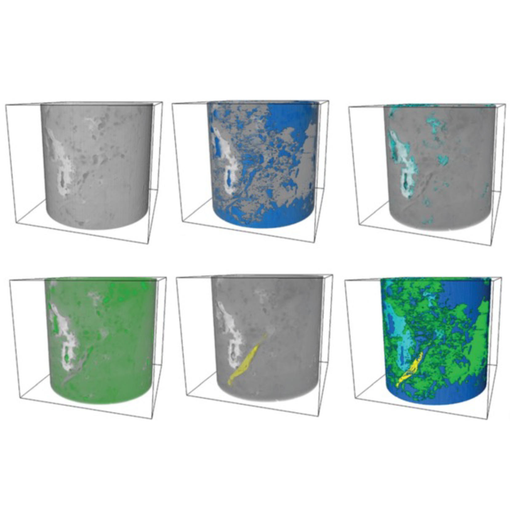
3D Microstructure of Soft Magnetic Elastomer Membrane
Soft magnetic elastomer membranes enable fast magnetic actuation under low fields. In our project, we… Read More
Events & Resources
News, Events and Resources from NXCT Partners
The thymus plays a crucial role in the human immune system. Thus, characterising its 3D structure is important to improve our understanding. Through the NXCT, we assessed the capabilities of x-ray CT for imaging the thymus. The results indicate that innovative CT techniques such as phase contrast are a powerful tool for our purposes.
| The thymus is an essential organ for the development of T-cells, which play an important role in the human immune response. Alterations in normal thymus function can result in severe illnesses of the immune system, so improving our knowledge about this organ can have an important impact in clinical cases. For this reason, there is scientific interest in engineering thymus-like microenvironments, which can illuminate how it works and, ultimately, support functional T-cell development.
Advanced imaging techniques are paramount for testing and validating these artificial thymus environments. The standard methods, such as electron microscopy and histology (see Fig. 1), provide important information but are constrained to 2D imaging and, in the latter case, require the destruction of the sample. In contrast, x-ray micro computed tomography (micro-CT) provides invaluable volumetric information on sample structure non-destructively. Traditional x-ray imaging techniques, which exploit the x-ray attenuation driven by the sample, tend to provide unsatisfactory contrast for soft-tissue samples such as the thymus. Instead, multi-contrast techniques like edge-illumination (EI) x-ray CT can improve soft-tissue contrast, particularly by exploiting the x-ray phase information. For this project, we imaged a critically-point dried native mouse thymus using EI CT. This was an important step in assessing the suitability of x-ray phase contrast micro-CT to the imaging of thymic tissue. The phase-contrast data indeed provided improved contrast between the cortex and the medulla compared to the attenuation (see Fig. 2). In addition, this work allowed to volumetrically visualise the mouse thymus and perform virtual dissections on it. Internal structures were observed across different planes of dissection, a feature unprecedented by other destructive imaging techniques. These data have provided a proof of concept for the feasibility of using multi-contrast x-ray micro-CT for characterising the 3D structure of the thymus. This is a new avenue of research and it will be very important in assessing the function of lab-engineered thymus micro-environments. It could provide a realistic pathway for better research on advanced therapies for thymus-related conditions. We will use these results to further explore the use of x-ray CT for thymus characterisation, possibly exploring efficient sampling strategies such as cycloidal CT.
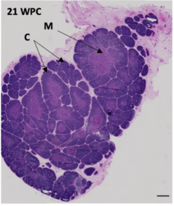 Figure 1. Histological section of a human foetal thymus (21 weeks post-conception), stained with the H&E protocol. The “M” and “C” correspond to “medulla” and “cortex” respectively, the same parts highlighted below in Fig. 2c. The final aim of this project will be to image the human thymus with high contrast and resolution. So far we have only obtained pilot x-ray CT data, but comparing our results in Fig. 2 to this histological slice shows that we can indeed see the morphological differences that we are interested in. Scale: 500 µm. 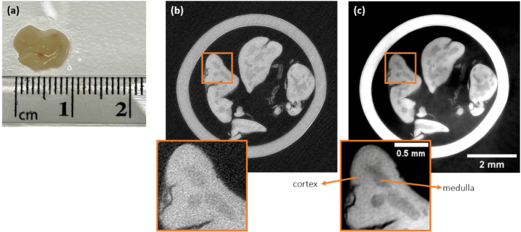 Figure 2. Phase-contrast EI provides an improvement in contrast between the different morphological parts of the thymus. In this project, a mouse thymus as seen in panel (a) was fixed and dehydrated, then imaged with EI. Panel (b) shows the resulting image using the traditional attenuation contrast, and panel (c) shows an image for the same slice using phase contrast. The improvement in telling apart the cortex and the medulla is visually apparent. This highlights the potential of EI for characterising the thymus in 3D.
|
Soft magnetic elastomer membranes enable fast magnetic actuation under low fields. In our project, we… Read More
Nowadays, the increasing capability of micro-manufacturing processes enables the manufacture of miniature products with extremely… Read More
Injection of CO2 into shale reservoirs to enhance gas recovery and simultaneously sequester greenhouse… Read More
