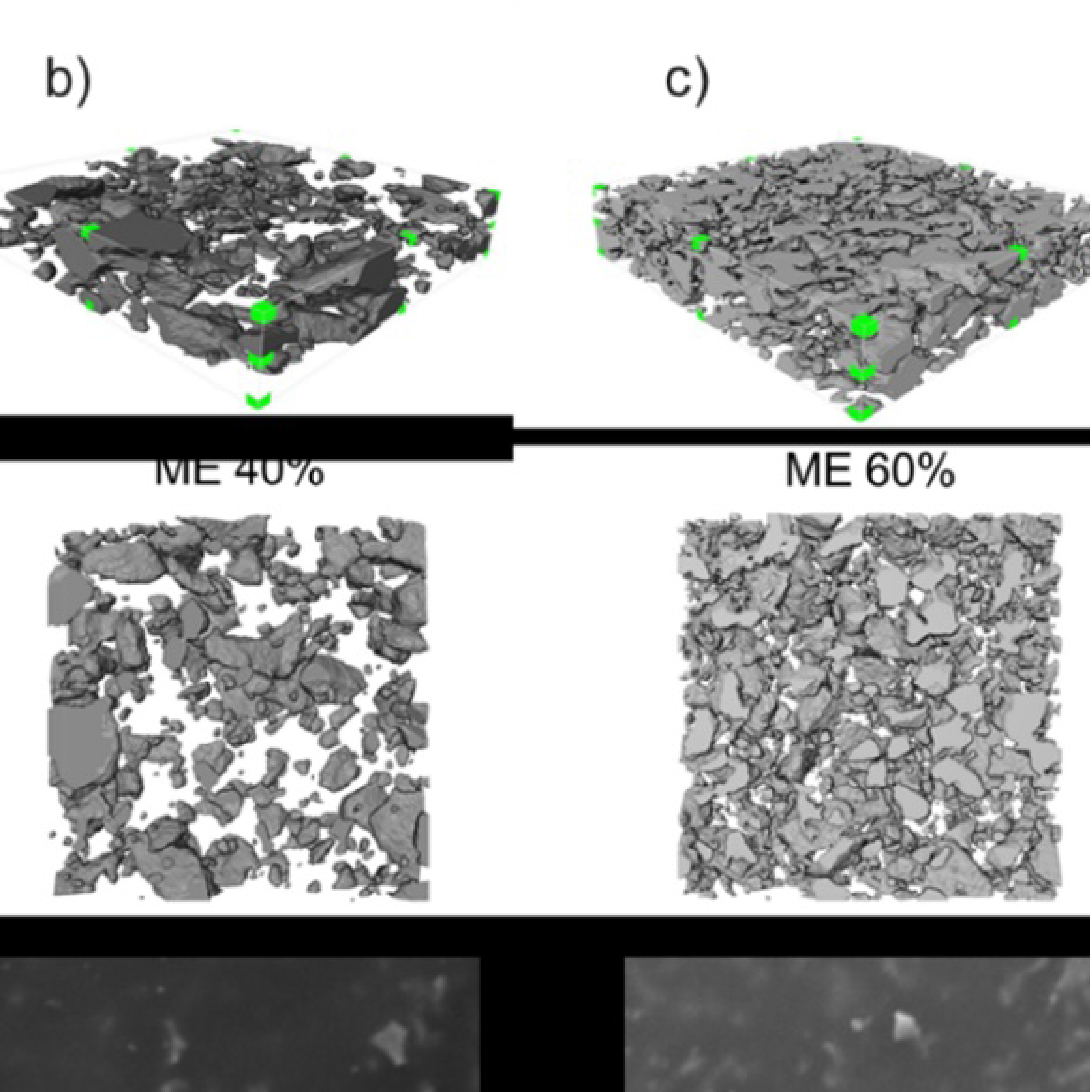
3D Microstructure of Soft Magnetic Elastomer Membrane
Soft magnetic elastomer membranes enable fast magnetic actuation under low fields. In our project, we… Read More
Events & Resources
News, Events and Resources from NXCT Partners
Using Micro-CT to evaluate the grain yield of different types of different genetic architectures of wheat spikes and corn cobs, allowing quantification and volumetric characterization of grains. This enables non-destructive, quantitative comparison of different wheat phenotypes and an exploration of analysing maize samples.
The work was carried out as a collaboration between the Centre of Imaging, Metrology, and Additive Technologies (CiMAT) and Prof. Jose Gutierrez-Marcos of the School of Life Sciences (SLS). The main motivation for the work was exploratory, as few studies investigating the potential for x-ray computed tomography for investigating crop yield currently exist.
Two main crop types were investigating to ascertain their potential for scanning, digitization, and quantification; individual wheat spikes and maize cobs. Both were scanned at the NXCT facility at the University of Warwick on their Tescan Unitom XL, prioritizing scan speed and contrast.
After preliminary scanning, it was determined that the wheat spikes showed high potential for quantification and comparison, with the maize samples also showing excellent contrast. As a result, a total of 30 wheat spikes were scanned using the same method and subsequently analysed.
Analysis was carried out by Dr. Paul Wilson at CiMAT, who extracted individual grains across all thirty samples in order to characterise a number of properties of each grain; 1) volume, 2) dimensions, 3) aspect ratio, 4) circularity, carried out using Avizo 2021.3.
X-ray computed tomography (CT) has the potential to revolutionise plant biology research by enabling non-destructive, high-resolution imaging of plant structures in three dimensions (3D). This technology has found broad applications in examining complex plant architectures, especially in studying inflorescences and seeds, where precise structural details are crucial for understanding developmental processes, functional morphology, and evolutionary biology. Traditional methods, such as histology, require slicing plant tissues, which can distort or destroy delicate structures. X-ray CT, in contrast, allows us to preserve the sample in its entirety while capturing intricate internal and external features. This is especially important for inflorescences, which are highly branched and can have small and fragile components. CT imaging allows the precise mapping of these structures, providing insights into how flowers and their arrangements develop, vary across species, and contribute to reproductive success.
In our study, X-ray CT is particularly valuable for exploring both macroscopic and microscopic structures. Inflorescences are often highly branched and intricate, with many developmental stages occurring simultaneously. Understanding the spatial arrangement of floral organs and their development is key to studying plant reproductive biology and evolution. X-ray CT will enable us to create detailed 3D models of inflorescences, capturing the relationships between different floral parts and the overall architecture without the need for dissection. This helps scientists understand the geometry and growth patterns of inflorescences in a way that traditional methods cannot match. Additionally, 3D CT models can be used to investigate how inflorescence architecture correlates with seed yield. The internal structure of seeds, including the arrangement of the embryo, endosperm, and seed coat, is fundamental to understanding seed development, dormancy, and germination processes. Traditional imaging methods often require cutting the seed, which can alter the native state of internal structures. X-ray CT, however, has allowed us to visualize these components in their intact state, providing accurate measurements of seed volume, density, and internal morphology. In addition to visualizing seed structure, X-ray CT can be used to examine how seeds respond to stresses like drought or temperature changes, revealing structural changes that are not easily detectable through other imaging techniques. This can help us to identify traits linked to seed resilience, which is important for crop improvement and conservation efforts.
High-throughput CT scanning can also be used to rapidly measure and compare morphological traits across many specimens, providing a wealth of data for genetic, developmental, and ecological studies. For both inflorescences and seeds, this capability enhances the ability to link phenotypic variation to underlying genetic and environmental factors.
Additional research is currently underway in collaboration with CiMAT, further exploring different phenotypes of wheat inflorescences and seeds. This has resulted in a successful BBSRC application entitled “A novel transcriptional pathway that controls axillary meristem induction in grasses: BB/X002535/1”.

Soft magnetic elastomer membranes enable fast magnetic actuation under low fields. In our project, we… Read More

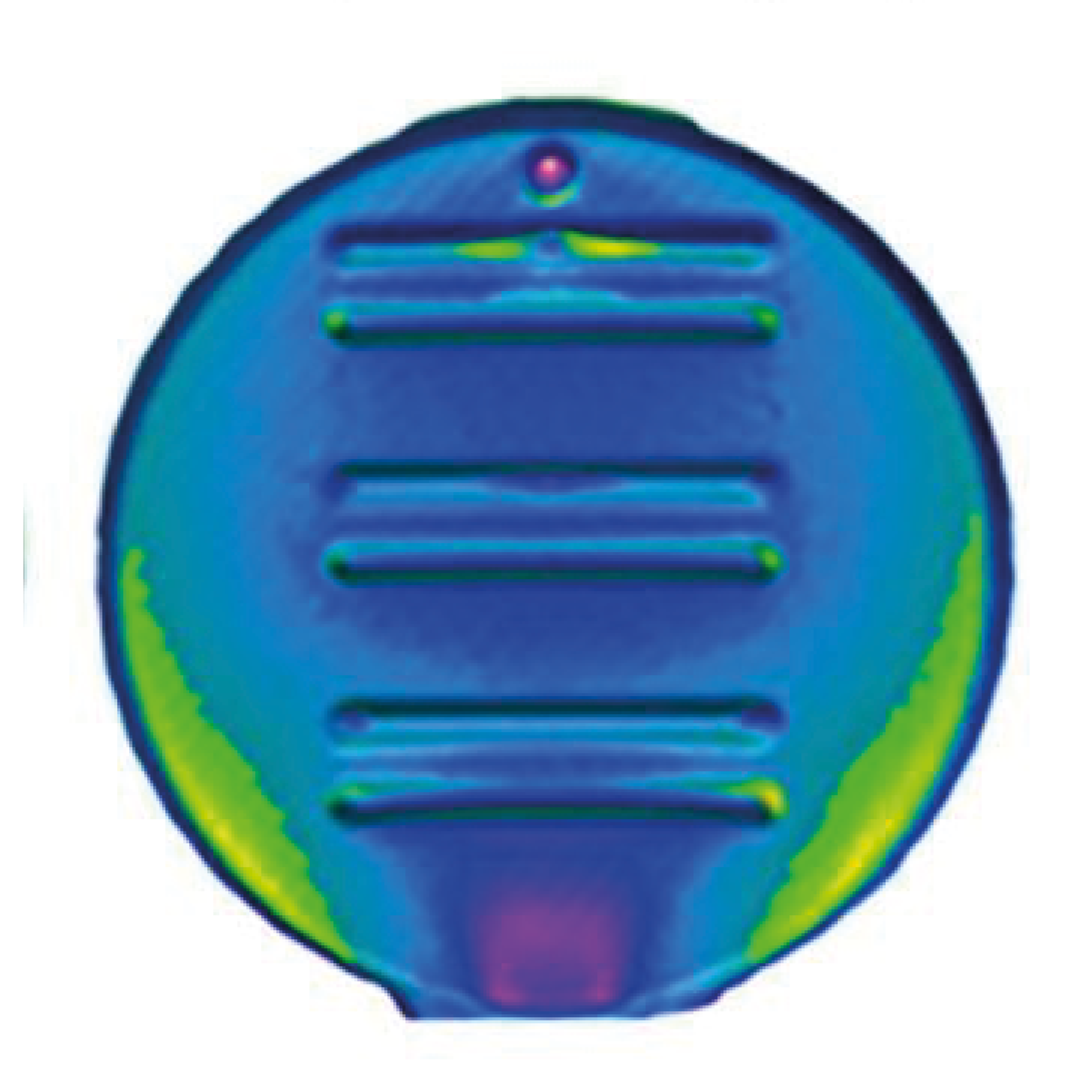
Nowadays, the increasing capability of micro-manufacturing processes enables the manufacture of miniature products with extremely… Read More

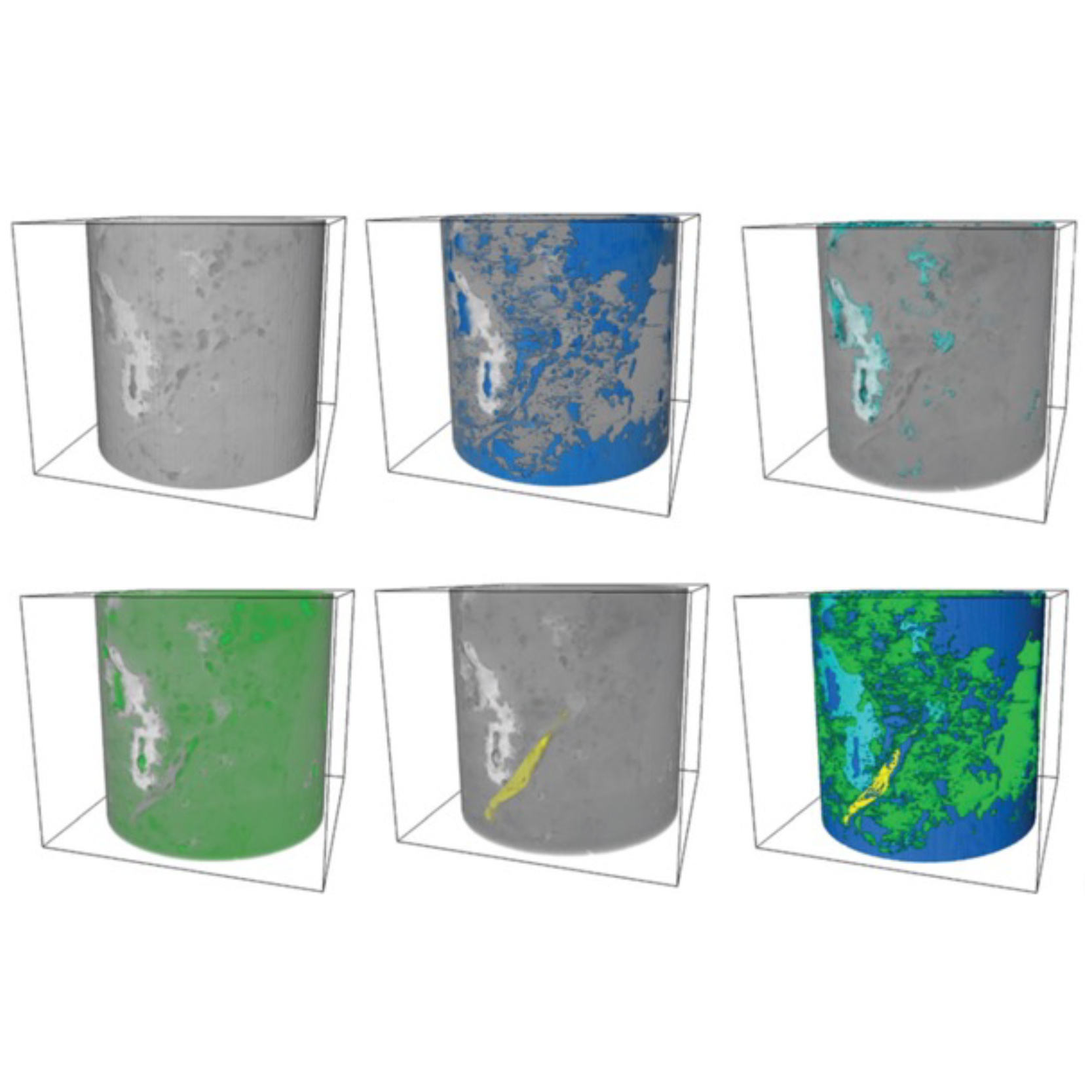
Injection of CO2 into shale reservoirs to enhance gas recovery and simultaneously sequester greenhouse… Read More