
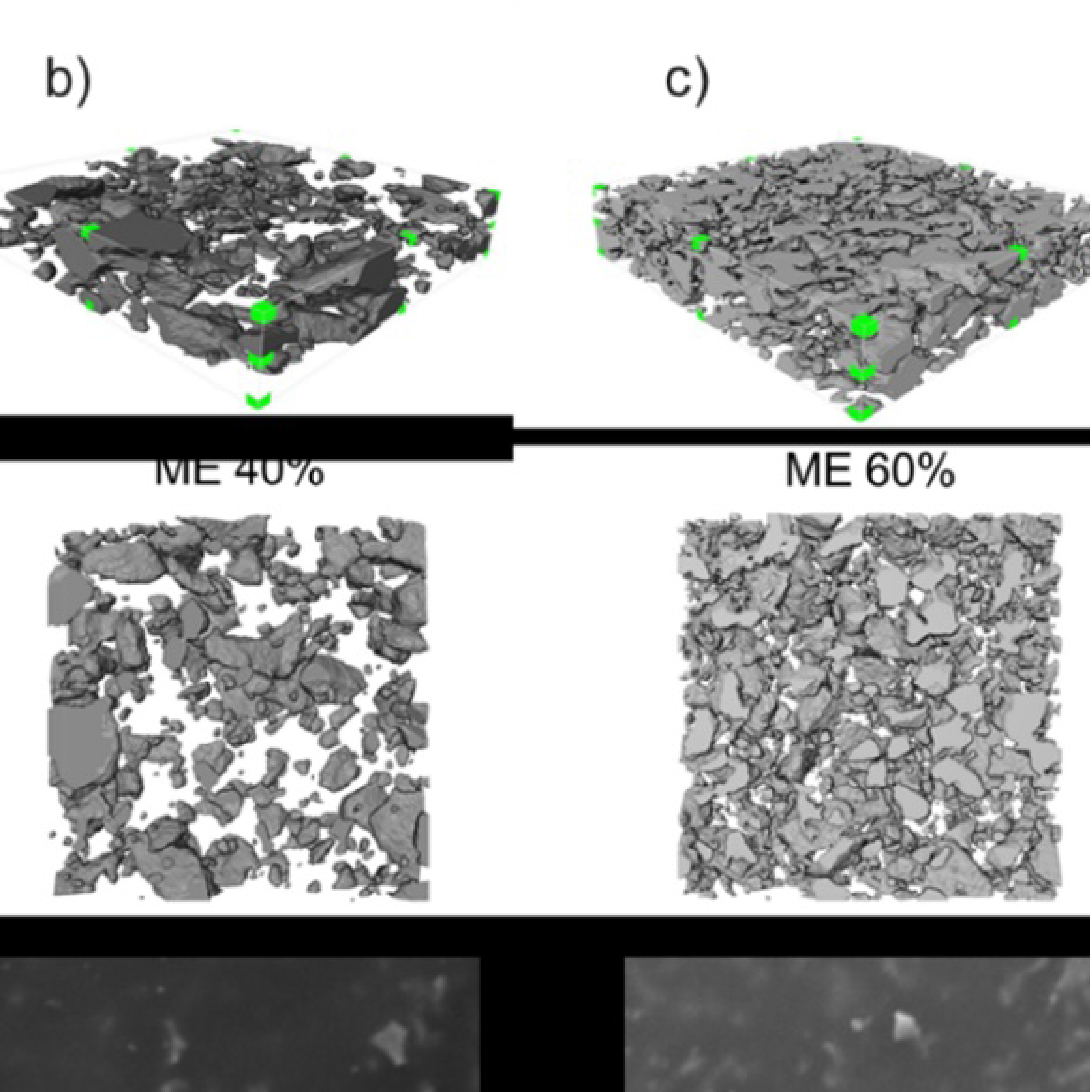
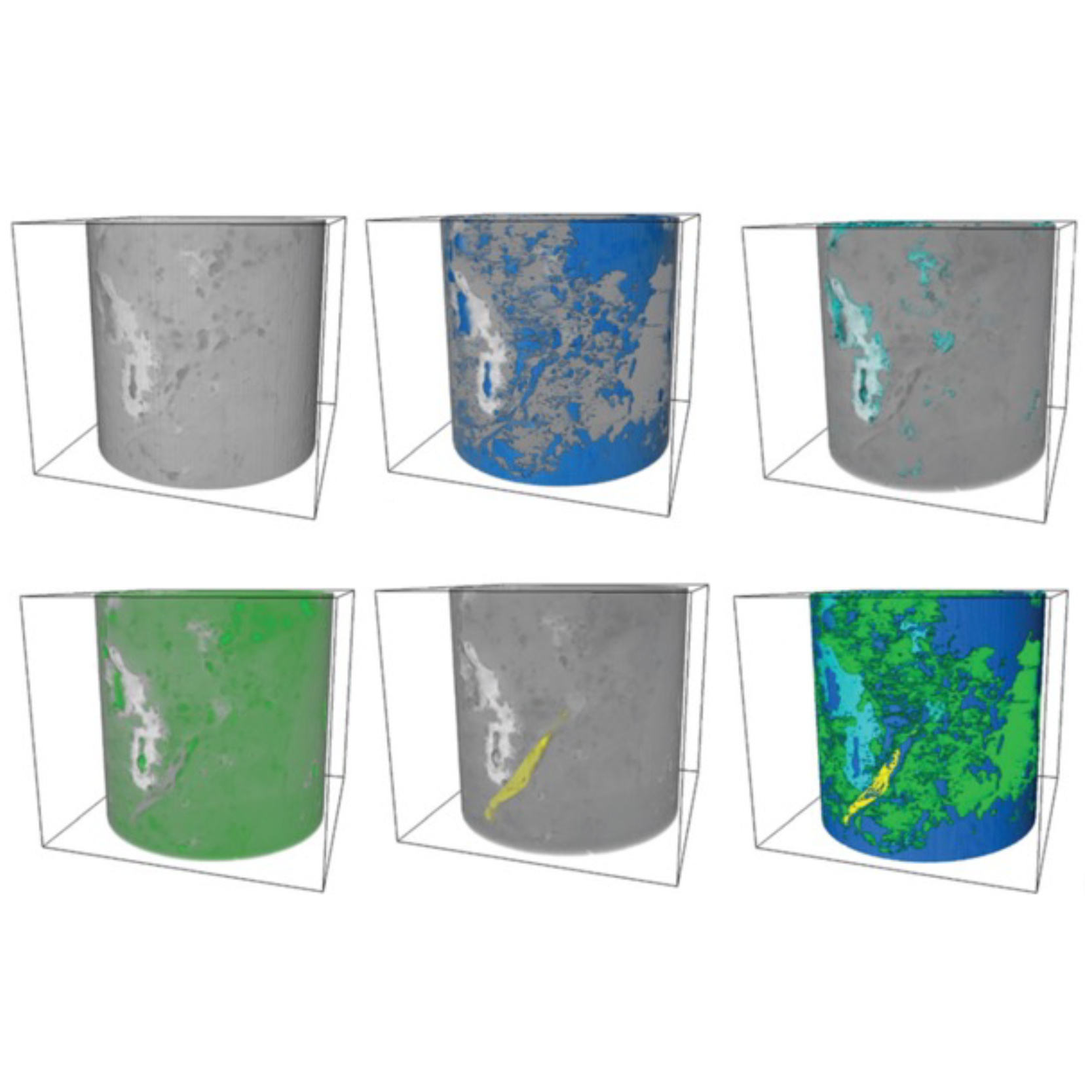
3D Microstructure of Soft Magnetic Elastomer Membrane
Soft magnetic elastomer membranes enable fast magnetic actuation under low fields. In our project, we… Read More
Events & Resources
News, Events and Resources from NXCT Partners
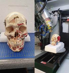
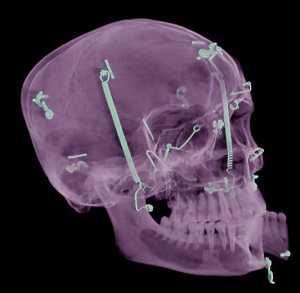
MicroCT has been used to create a virtual 3D model of a precious and delicate antique anatomical preparation of a dissected and annotated human skull human skull. The digital version will be used to create a 3D-printed and student-proof copy for teaching and outreach purposes.
The Centre for Learning Anatomical Sciences (CLAS) in Medicine at Southampton University trains medical students in anatomy and surgery. It holds a museum collection of normal and pathological specimens including a human skull that has been sliced in various planes, articulated and painted to reveal and annotate structures not normally seen. The skull has significant value as a teaching aid but is too fragile to actually be used. A chance conversation between staff of CLAS and those of the Biomedical Imaging Unit (BIU), Medicine’s microscopical imaging facility, developed into a project to try to create a student-proof replica of the skull.
A 3D print requires a surface mesh from which to generate the printer files, and microCT was the obvious choice, with the additional benefit of revealing internal detail not visible in the original preparation.
The BIU works in close collaboration with Southampton’s μ-VIS X ray Imaging Centre, part of the NXCT consortium and we applied for free beam time to generate the scans and for initial data processing. The skull was scanned on their custom Nikon XTH225ST system at Southampton General Hospital by Drs. Orestes Katsamenis and Stephanie Robinson.
Because of the hard contrast between the metal fittings and bone, there is inevitable shadowing in the data set but this will not affect the processing now needed to generate the mesh. That process, optimising the print files and subsequent printing and painting will all take time, but, thanks to NXCT and μ-VIS, the journey is underway. The potential of microCT to help create durable replicas of rare and delicate anatomical specimens is confirmed and advances in soft tissue CT imaging can extend this to other historic anatomical material. All parts of this collaboration are being undertaken in compliance with the Human Tissue Act.
“The results thus far have exceeded all expectations. I am impressed by the depth and detail revealed in by the scanning process. The potential for further integration into education are very exciting. CLAS looks forward to any future exchange of ideas that would enhance the educational experience for our students.”
Mr Jed Clampitt, Anatomical Sciences Laboratory Technician, Centre for Learning Anatomical Sciences, Faculty of Medicine, University of Southampton.
Dr. Dave Johnston, Facility Manager, Biomedical Imaging Unit, Faculty of Medicine, University of Southampton.
Collaborators
Dr Orestis Katsamenis and Dr Stephanie Robinson, μ-VIS X ray Imaging Centre, School of Engineering, University of Southampton
Fig. 1: The specimen being prepared for scanning (image: Orestis Katsamenis)
Fig. 2: Virtual 3D model from the CT dataset (image: Orestis Katsamenis)
Soft magnetic elastomer membranes enable fast magnetic actuation under low fields. In our project, we… Read More
Nowadays, the increasing capability of micro-manufacturing processes enables the manufacture of miniature products with extremely… Read More
Injection of CO2 into shale reservoirs to enhance gas recovery and simultaneously sequester greenhouse… Read More
