Affiliates
-
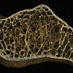
- 1) Human bone from the Mary Rose remains. Image courtesy of Dr S Aldridge
-
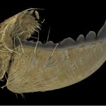
- 2) Zinc-tipped leafcutter ant mandible. Image courtesy of Prof. R Johnston
-
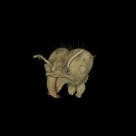
- 3) Leafcutter ant head. Zinc-rich region highlighted red. Image courtesy of Prof. R Johnston
-
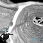
- 4) Virtual Reality headset view inside microCT data of a dragonfly wing vein
IM3AGES at Strathclyde
The Im3ages Facility at Strathclyde Hosts a three highly complementary X-ray tomography systems, a dual head Nikon XTH 180/225, a Zeiss Versa 730 XRM (with DCT), and a Tescan dynaton. These systems extend national capability and capacity, and allows Im3ages to offer high resolution, high speed, high throughput and large sample lab XCT to our users and to the NXCT. Our users generally come from the materials, engineering and manufacturing, geology and environmental science, bioscience and agriculture, and cultural heritage sectors; but we have scanned all sorts of objects for research and industry partners across the UK.
As our Dynatom system has the option of a static or rotating sample during imaging and the ability to collect scans in less than 1 minute, Im3ages has a strong focus on in situ experiments. We host a range of experimental cells that can be used in Im3ages scanners, or at other NXCT facilities. These include mechanical testing with the Deben CT5000 cryo and high temperature load cells, or 10kKN open frame rig (all of which can accommodate saturated samples), flow and porous media experiments where we can get to reservoir pressure and temperature conditions, high temperature furnaces, and rheological testing.
AIM at Swansea
The Advanced Imaging of Materials (AIM) Core Facility at Swansea University contributes complementary X-ray CT capability to NXCT through its Zeiss Xradia Versa 520 and Nikon XTH 225 systems, enabling high-resolution and large-sample 3D imaging across materials, engineering, energy, cultural heritage, and bio-inspired research. AIM brings strong experience in in situ and operando X-ray CT, including mechanical testing using the Deben HT5000 and Bruker Hysitron Intraspect nanoindentation platforms, as well as temperature-controlled studies using a Deben cooling stage. A key contribution to NXCT is AIM’s integrated correlative imaging approach—linking X-ray CT with SEM, FIB-SEM and spectroscopy—alongside advanced VR-based volumetric analysis, which enables immersive, full-3D interrogation of CT datasets and supports collaborative analysis, training and engagement.
STFC
Professor Frank Vidal leads the R&D activities related to X-ray simulations at Scientific Computing – Science and Technology Facilities Council (STFC) [sc.stfc.ac.uk]. He develops and maintains the award-winning open-source programming framework called gVirtualXray (gVXR) [gvirtualxray.sourceforge.io]. It is used to simulate realistic X-ray images on GPU. It has been used in a wide range of applications, including real-time medical simulators, proposing a new densitometric radiographic modality in clinical imaging, studying noise removal techniques in fluoroscopy, teaching particle physics and X-ray imaging to undergraduate students in engineering, and XCT to masters students, predicting image quality and artifacts in material science, etc. gVXR has also been used to produce a high number of realistic simulated images in optimisation problems and to train machine learning algorithms, in particular for image segmentation and object detection. More recently, digital twins of actual XCT devices were integrated in gVXR. A modern Web-UI, called WebCT [webct.io], is also available. They can be used to plan and optimise the scanning of a given sample using a particular device. His colleagues at Scientific Computing [sc.stfc.ac.uk] develop CIL [ccpi.ac.uk] for CT reconstruction and iDVC [tomographicimaging.github.io] for Digital Volume Correlation analysis.